Respiratory Emergencies: The First 60 Seconds Needle
Dr Chris Parratt MRCVS
Parrattchris@hotmail.com
Respiratory Emergencies: The First 60 Seconds
Breed
Common Aetiology
Notes
Cavalier King Charles Spaniel
Congestive Heart Failure
Usually mitral valve disease
Yorkshire Terrier, Pomeranian
Tracheal collapse
Benefit from sedative
Irish Wolfhounds
Congestive Heart Failure, Aspiration
CHF usually due to Dilated
pneumonia
cardiomyopathy (DCM)
Brachycephalic Upper Airway Syndrome
Check temperature, often
(BUAS)
hyperthermic & require cooling
Congestive Heart Failure
Usually DCM. Risk of sudden
Pug, Bulldog
Doberman Pinscer, Boxer, Great Dane
cardiac death
Young cats
Older cats
Infection (pyothorax?), Congestive heart
Check temperature: may be fever
failure
with infection
Congestive heart failure, neoplasia
Often pleural effusion – needle
thoracentesis.
Oriental cats (eg Siamese, Bengal)
Allergic Airway Disease
Expiratory effort. Often cough.
Table 1: Cardiorespiratory breed predispositions encountered in the authors’ experience.
Disease Localisation
Upper Respiratory
Breathing Observation
Noisey (Stridor and
stertor)
Rapid and shallow.
Examples
Laryngeal paralysis, BUAS
Comments
Often hyperthermic –
require cooling
Lung Parenchyma
Pulmonary oedema,
Crackles or wheezes on
pneumonia, contusions
auscultation?
Small airway
Expiratory wheeze
Allergic airway disease
Oriental cats predisposed
Pleural space
Paradoxical Breathing
Pleural effusion,
Heart and lungs
pneumothorax,
inaudible/muffled on
diaphragmatic hernia
auscultation
Table 2: Localisation of respiratory pathology. More than one disease process may be present concurrently, making
interpretation using this method difficult.
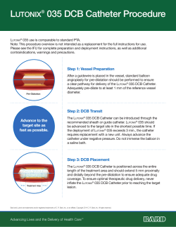
Needle Thoracocentesis
Equipment:
60ml syringe (or similar)
3-way tap
Butterfly needle (or ‘over the needle’ catheter &
extension tubing)
Technique: Local anaesthesia is not required. In emergency
situations, aseptic preparation may be detrimental and the skin
simply prepped with an alcohol solution.
The site of insertion is selected: 8-11th intercostal space. Avoid the
caudal aspect of the rib, to avoid the intercostal neurovascular
bundle.
If pneumothorax is suspected, insert in the dorsal third of the
thorax. If Blood (or other fluid) is suspected, insert the needle in
the ventral third of the thorax.
The needle should be carefully advanced, whilst the syringe
plunger is simultaneously withdrawn, inducing a negative
pressure. As soon as the thoracic cavity is entered, the negative
pressure is lost, and the pleural contents start to become
aspirated.
To reduce the risk of pulmonary trauma, the needle should be
promptly oriented parallel with the thoracic wall.
Dr Chris Parratt MRCVS
Parrattchris@hotmail.com
Emergency Tracheotomy (Reproduced with permission from Ed Cooper)
Patient must be unconcious or anaesthetised. Only effective if tracheotomy distal to obstruction.
Approximate inhaled O2 (%)
Oxygen cage
21-60
Flow By Oxygen
24-45
Face Mask
35-45
Unilateral Nasal Catheter
30-50
Bilateral Nasal Catheter
30-70%
GA & Intubation
21-100
Table 3: Approximate inhaled oxygen concentration (FiO2) with different methods of oxygen
delivery.
Nasal Oxygen Catheter
Equipment:
2% Lignocaine (without
adrenaline)
KY Jelly
Flexible catheter (Nasal catheter,
urinary catheter, other soft tubing)
Technique:
- Measure from nostril to lateral canthus of
eye
- Instill lignocaine into nostril (5mls Dogs,
1ml Cats)
- Lubricate Catheter with KY jelly
- Insert catheter in ventro-medial direction
- Secure in place with butterfly strips
(superglue or skin stapler)
- Connect to Oxygen (Humidified?)
- Bilateral catheters can be placed to
increase inspired oxygen concentration.
© Copyright 2025


