Lisfranc Injuries and Surgical Treatment Jacques Lisfranc Lisfranc Definition
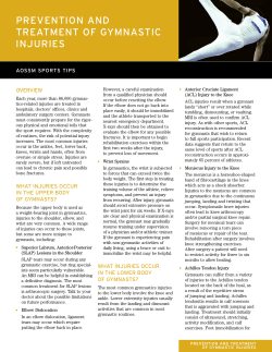
PAOS 2012 CME Conference ~ Hilton Head Island, SC ~ October 22 - 26, 2012 Jacques Lisfranc Lisfranc Injuries and Surgical Treatment Bruce Cohen MD OrthoCarolina Foot and Ankle Institute Charlotte NC • French surgeon in Napoleon’s army • Described a midfoot amputation for gangrene Nouvelle methode operatoire pour l’amputation partielle du pied par son Articulation tarso-metatarsienne: 1815 Lisfranc Definition • Any bony or ligamentous injury that involves the TMT joints • Multiple variants Anatomy Anatomy • Very rigid: bony architecture Mechanisms for Injury Direct • Very rigid: ligaments –Lisfranc ligament most important stabilizer Lisfranc Injuries and Surgical Treatment • Plantar force • Atypical fracture patterns • Primarily bony component 1 PAOS 2012 CME Conference ~ Hilton Head Island, SC ~ October 22 - 26, 2012 Mechanism for Injury Mechanism for Injury Indirect Indirect • Common sports injury • More common – Axial load to the back of the heel with foot fixed to the ground – Athletes • Axial load • Often purely ligamentous Wide Variety of Patterns Possible! Diagnosis Exam • Indirect types may be subtle • Painful WB • Swelling and point tenderness Radiographs Diagnosis • • • • • • Neurovascular compromise • Compartment syndrome Lisfranc Injuries and Surgical Treatment Mandatory part of the work-up AP/lateral/oblique of foot Weightbearing is critical! Consider contralateral films 20% of these injuries are missed on initial evaluation 2 PAOS 2012 CME Conference ~ Hilton Head Island, SC ~ October 22 - 26, 2012 AP View • • • • 1st TMT joint 2nd TMT joint 1-2 interspace Naviculocuneiform Oblique View • 2nd TMT Joint • 3th TMT Joint • 4th TMT Joint Radiographs Lateral View “fleck” sign • 1st/2nd TMT Joints Radiographs • Subtle signs – MT neck fracture – MP dislocation – “Fleck sign” – Compression fracture of cuboid Lisfranc Injuries and Surgical Treatment • Standing AP is good stress test! • Consider contralateral xray Radiographs • Subtle signs – MT neck fracture – MP dislocation – “Fleck sign” – Compression fracture of cuboid 3 PAOS 2012 CME Conference ~ Hilton Head Island, SC ~ October 22 - 26, 2012 Radiographs Radiographs • Standing AP is good stress test! • Subtle signs – Beware of the proximal variant! – Increasing incidence in NFL • Hammit/Anderso n, TFAS ‘05 Non-weightbearing Weightbearing Proximal variant • Results in an unstable first ray difficulty with push-off Stress Testing Formal stress testing • Requires anesthesia, flouroscopy • Maneuver – Adduction-pronation Diagnostic Evaluation Stress Testing CT • Unusual fx patterns • May help guide treatment • Confirm nondisplaced (nonop) Lisfranc Injuries and Surgical Treatment 4 PAOS 2012 CME Conference ~ Hilton Head Island, SC ~ October 22 - 26, 2012 Diagnostic Evaluation MRI • Helpful if a vague presentation; “sprain” • May assist in treatment and prognosis Nonoperative Treatment • Stable injury pattern (non-displaced) • 6-8 weeks protected weightbearing – Short-leg cast – Walker boot • Repeat radiographs • Expect 3-4 month recovery Surgical Timing • If excessive swelling, consider delaying surgery 10-14 days • All severe dislocations should be reduced urgently • Compartment syndrome – operate acutely Lisfranc Injuries and Surgical Treatment Treatment Goal • Obtain/maintain precise anatomic reduction • Preserve a stable, plantigrade foot Surgical Indications • All unstable injury patterns –Tarsometatarsal joints –Intercuneiform joints • Open fracture • Compartment syndrome • NV compromise Recommendations for Fixation • Treat individually – Pain? – Can not push-off? – Progressive diastasis? – Unstable pattern confirmed by stress? Weight-bearing 5 PAOS 2012 CME Conference ~ Hilton Head Island, SC ~ October 22 - 26, 2012 Surgical Technique • • Open reduction Medial column (TMT 1-3) – Screw fixation including “homerun” screw – Option: bridge plate for TMT joints • Lateral column (TMT 4-5) – Pin fixation • Lateral crush (i.e. cuboid) – Consider ex-fix Surgical Technique - Fixation Since injury primarily ligamentous… • Takes months to heal • Use “rigid” internal fixation – Screws = gold standard • Avoid cannulated screws – risk for breakage Surgical Technique Surgical Technique • Open reduction – Removes debris • Leave soft tissue/ligaments – Can assess cartilage – Intercuneiform of other subtle areas of instability? – Confirms anatomic reduction Surgical Technique • Bridge plates becoming popular as an option to screws - avoids articular cartilage damage with no loss of rigidity – Ligamentous Lisfranc Joint Injuries: A Biomechanical Comparison of Dorsal Plate and Transarticular Screw Fixation – Frank G. Alberta, M.D; Michael S. Aronow, M.D; Mauricio Barrero, M.S; Vilmaris Diaz-Doran, B.S; Raymond J. Sullivan, M.D; Douglas J. Adams, Ph.D Farmington, CT » Foot & Ankle International, Vol. 26, No. 6, June 2005 Surgical Technique • Bridge plates Comminuted fracture • Consider “bridge” plate Lisfranc Injuries and Surgical Treatment – Can use on 1st and 2nd TMT joints – Locking screws beneficial 6 PAOS 2012 CME Conference ~ Hilton Head Island, SC ~ October 22 - 26, 2012 Case Example Case Example 40 yo involved in Motorcycle crash Case Example Surgical Technique Lisfranc surgical system • Reduction devices • Solid screws – Cannulated assisted 50 yo involved in Fall Surgical Technique Postoperative • SLC, NWB x 4-6 wks • Boot, NWB X 4-6 wks • Pin removal (if used) at 6 wks • Screw/plate removal 4-6 months – Ligament injuries require longer period of protection Lisfranc Injuries and Surgical Treatment 7 PAOS 2012 CME Conference ~ Hilton Head Island, SC ~ October 22 - 26, 2012 Screw/Plate Removal • Controversial • Remove all lateral column (TMT 4-5) fixation • Prefer to remove transarticular TMT 1-3 hardware 1° Arthrodesis • Indications? – Late presentation (8-12 wks) – Severe articular damage – Risk for malunion and nonunion – High energy?? 1° Arthrodesis • Treatment of primarily ligamentous Lisfranc joint injuries: primary arthrodesis compared with open reduction and internal fixation. Surgical technique. Coetzee JC, Ly TV JBJS 2007 – – – – Prospective RCT 41 pts Ligamentous injury Better short term outcome with primary fusion 1° Arthrodesis • Open reduction internal fixation versus primary arthrodesis for lisfranc injuries: a prospective randomized study. Henning JA, Jones CB, Sietsma DL, Bohay DR, Anderson JG Foot and Ankle Int 2009 – 32 pts – Randomized – Lower subsequent procedures with arthrodesis – Biased study – Still reasonable to do arthrodesis Prognosis • Expect long rehab (> 1 year) – Midfoot pain/stiffness for average 1.3 years postop (Brunet) • Ultimate outcome related to adequacy of reduction and severity of initial injury Lisfranc Injuries and Surgical Treatment Additional Cases 8 PAOS 2012 CME Conference ~ Hilton Head Island, SC ~ October 22 - 26, 2012 Case GS • 24 y/o basketball player with twisting injury to foot while rebounding • Midfoot pain/swelling • Unable to run Case GS Case GS • Xrays reportedly normal • Crutches/NWB x 3 weeks, but persistent pain • Consult: standing Xrays obtained Case GS • Tender midfoot • No instability • What test would you order? –CT Scan –Bone Scan –MRI Case GS Lisfranc Injuries and Surgical Treatment Case GS 9 PAOS 2012 CME Conference ~ Hilton Head Island, SC ~ October 22 - 26, 2012 Case GS • Lisfranc sprain – no instability • How should we treat him? • What if he’s not better after 3 months? • 6 months? One year? Case DC • Tender midfoot but minimal swelling • Pain with medial-lateral compression of foot Case DC Case DC • 25 y/o professional quarterback • Twisting injury while avoiding a sack • Took himself out after one play with painful weightbearing Case DC • Standing Xrays – Concerned? Take Home Points • These are easily missed • Standing Xrays – Look at n-c joint – Get comparison views with other side – Have a high index of suspicion • Need weight bearing films – If they can’t, they need repeat exam in a week • If concerned get follow-up sooner than later • Advanced imaging is helpful – CT with fractures – MRI with ligament/sprains Lisfranc Injuries and Surgical Treatment 10 PAOS 2012 CME Conference ~ Hilton Head Island, SC ~ October 22 - 26, 2012 Thank You Lisfranc Injuries and Surgical Treatment 11
© Copyright 2025

