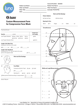
Customized immobilization face masks for radiotherapy using
Customized immobilization face masks for radiotherapy using Incremental Sheet Forming
Treatment of head and neck tumours requires radiotherapy with high precision localized to the diseased area
with the tumour [1]. Missing the desired area can lead to increased doses to sensitive body parts such as the eye
or the spinal cord [1, 2]. Present immobilization systems for head fixation typically use masks made of PVC or
thermos-plastic material such as Orfit masks [3]. A study conducted by Weltens et al. [1] showed errors larger
than 4 mm in 6.2% cases where thermoplastic masks were used [1]. Furthermore, closed form, full head masks
have a setup uncertainty of 2-3 mm and force patients to keep the eyes and mouth closed during daily treatment
and may be uncomfortable for patients with claustrophobia [4].
To overcome the issues associated with the current fixation techniques in immobilization for head and neck
radiotherapy, this project will seek to develop customized immobilization face masks using digital images of the
patient and a flexible sheet prototyping process, Incremental Sheet Forming (ISF), available in the Faculty of
Engineering.
The digital images of the patient will be processed to generate 3D point clouds. The reconstruction from images
to point clouds will be based on identifying local features in the images which will be extracted and matched
between images. The matches will be used to compute relative poses between image pairs and to triangulate a
sparse reconstruction, following the work done in developing the web-based service Arc3D [5]. In addition, the
accuracy of the generated point clouds will be evaluated by comparison with laser scanner based reconstruction.
Once the algorithms for reconstruction from images are well validated, these point clouds will then be meshed
using meshing routines such as Marching Cubes and the mesh will be used to generate toolpaths for incremental
forming within a software tool created specifically for this project.
The manufacturing challenges within this project will involve assessing the suitability of specific alloys for
making these masks and developing advanced toolpath strategies to form masks for a wide range of human
faces. Typically, parts made using ISF tend to fail as the wall angle of the formed part exceeds a limit dependent
on the material type and sheet thickness. In a face mask, failure is expected in the chin and nose regions where
the wall angles are typically high. Optimized toolpaths that take into account part failure limits and inaccuracies
in the formed part due to material springback and feature behaviour will be developed within the framework of
this project. Specifically, the suitability of multi-step morphing strategies and use of feature based material
specific generic compensation functions will be evaluated in detail.
The face masks formed in this project will be sent to collaborating clinics for evaluation with respect to
alignment and positioning, patient comfort etc. Feedback will be used to calibrate and validate the technique for
regular clinical use. This project will require interdisciplinary work involving close collaboration between
manufacturing engineering, computer science and medical science.
References
[1] C Weltens, K Kesteloot, G Vandevelde, WV Bogaert. Comparison of plastic and orfit masks for patient
head fixation during radiotherapy: precision and costs (1995). International Journal of Radiation Oncology
Biology Physics. Vol. 33, No. 2, pp. 499-507.
[2] CF Hess, R-D Kortmann, R Jany, A Hamberger, M Bamberg. Accuracy of field alignment in
radiotherapy of head and neck cancer utilizing individualized face mask immobilization: a retrospective
analysis of clinical practice (1995). Radiotherapy and Oncology. Vol. 34. pp. 69-72.
[3] Orfit Industries, http://www.orfit.com/, Last accessed 18th February, 2015
[4] G Li, M Lovelock, J Mechalakos, S Rao, C Della-Biancia, H Amols, N Lee. Migration from full-head
mask to “open-face” mask for immobilization of patients with head and neck cancer. Journal of Applied
Clinical Medical Physics (2013). Vol. 14. pp. 243-254
[5] Maarten Vergauwen and Luc Van Gool, "Web-Based 3D Reconstruction Service", Machine Vision
Applications, 17, pp. 411-426, 2006
Supervisors
Faculty of Engineering: Dr. Amar Kumar Behera, Research Fellow, Materials, Mechanics and Structures
Faculty of Science: Dr. Andrew French, Assistant Professor, Computer Science
Dr. Hengan Ou, Associate Professor, Faculty of Engineering
Collaborators
*Nottingham University Hospitals Trust: Russell Hart, Radiotherapy Services Manager
*Contacts were made with Russell Hart of NUH, Apollo Hospitals, India, Fortis Memorial Research Institute and a number
of radiotherapy clinics already. Confirmation of collaboration and use of research output is awaited in some cases.
© Copyright 2025


