
Elucidation of the action mechanism of erxian decoction, Title
Title
Author(s)
Elucidation of the action mechanism of erxian decoction,
a Chinese medicinal formula for menopause:
frompharmacological approach to analytical approach
Cheung, Ho-pan.; 張浩斌.
Citation
Issue Date
URL
Rights
2013
http://hdl.handle.net/10722/188299
The author retains all proprietary rights, (such as patent
rights) and the right to use in future works.
Abstract of thesis entitles
Elucidation of
the Action Mechanism of Erxian Decoction,
a Chinese Medicinal Formula for Menopause:
from Pharmacological Approach to Analytical
Approach
Submitted by
CHEUNG Ho Pan
(張浩斌)
for the degree of Master of Philosophy
at The University of Hong Kong
in Mar 2013
As the aging in reproductive system proceeds, females will eventually enter the
period of menopause, during which a series of physiological changes occurs. The
decline of estrogen level during menopausal transition is thought to associate with
various menopausal symptoms. Although hormone replacement therapy can be
adopted to deal with the estrogen-deficient state, side effects such as cancer risk
cannot be overlooked.
Alternatively, Erxian Decoction (EXD), a Chinese medicine formula for treating
menopausal symptoms has been used clinically for more than 60 years without
adverse effects reported. Some pharmacological properties of EXD have been
reported in previous research, which are thought to be contributed by its multiple
bioactive components. Thus in the present study, the pharmacological properties
of EXD have been further evaluated. The drug compatibility of Traditional
Chinese Medicine (TCM) formula, EXD, was also demonstrated. At last, a novel
approach for identification of bioactive components from Chinese medicine
formula was introduced using EXD as study model.
To evaluate the multiple pharmacological properties of EXD, proteins involved in
steroidogenesis in ovaries of aged female rats were measured by immunoblotting
analysis. On top of that, serum lipid profiles and the related proteins were
determined by colorimetric assay and immunoblotting analysis respectively. Also,
anti-osteoporotic properties and drug compatibility of EXD were evaluated by in
vitro methods such as proliferation assay, osteoclast differentiation assay, ELISA
assay or immunoblotting analysis. Lastly, a novel approach for identification of
bioactive components in relation to the subsequent bioactivity from traditional
Chinese medicinal formula was introduced using HPLC profiles.
From the results, it was demonstrated that EXD can modulate steroidogenesis in
aged female rat model at least through up-regulation of ovarian aromatase, protein
kinase B and estrogen receptor beta at protein level. Besides, EXD also exerts
antihyperlipidemic effects in aged female rats as reflected from the decreased
serum total cholesterol and LDL-cholesterol levels via regulation of HMG CoA
reductase and LDL-receptor, the key proteins for cholesterol synthesis and
LDL-cholesterol clearance. In vitro study has also demonstrated the
anti-osteoporotic properties of EXD through stimulation of osteoblast
proliferation and inhibition of proliferation and differentiation of osteoclast
precursor cells. The
NFATc1 proteins, a
categories according
contributing to the
elucidated.
later was proved to be mediated by down-regulation of
key protein for osteoclastogenesis. The roles of the drugs
to the drug compatibility of traditional Chinese medicine
optimal anti-osteoporotic properties of EXD were also
Since the diverse pharmacological properties of a Chinese medicinal formula are
often the results of the effects of complex bioactive constituents in the extract, yet
identification of the bioactive components has been a tedious task. Thus in the last
part of the study, a novel approach for identification of bioactive component from
Chinese medicinal formula has been developed. By comparing the HPLC profiles
of EXD extracted by different decoction method in relation to their
pharmacological properties, six bioactive chemicals were successfully identified
which may contribute to the stimulatory effect of EXD on ovarian aromatase and
hepatic catalase expression.
Elucidation of
the Action Mechanism of Erxian Decoction,
a Chinese Medicinal Formula for Menopause:
from Pharmacological Approach to Analytical
Approach
by
CHEUNG Ho Pan
(張浩斌)
A thesis submitted in partial fulfillment of the requirements for
the Degree of Master of Philosophy
at The University of Hong Kong
Mar 2013
Declaration
I declare that the thesis and the research work thereof represents my own work,
except where due acknowledgement is made, and that it has not been previously
included in a thesis, dissertation or report submitted to this University or to any
other institution for a degree, diploma or other qualifications.
Signed ____________________________
CHEUNG Ho Pan
i
Acknowledgement
I would like to express my gratitude to my principle supervisor Dr. Sze Cho Wing
Stephen, and my co-supervisors Prof. Tong Yao, Dr. Zhang Yanbo, Dr. Rong Jian
Hui, and Dr. Lee Kai Fai Calvin for their supervision, constant support and
encouragement throughout the course of the postgraduate study, as well as for the
advices and guidance on the preparation of thesis.
My gratitude is extended to the laboratory colleagues and team members
including but not limited to Mr. Wong Kam Lok, Mr. Wong Yiu Long, Mr. Ip
Chun Wai, Ms. Annballaw Leigh, Dr. Chu Shihng Meir Ellie, Ms. Lu Jia and Mr.
Zhang Liang for their helpful sharing and support. The technical support from Mr.
Wong Hei Kiu, Ms. Lee Wai Sin and Mr. Tsang Kam Wah is greatly appreciated
as well.
ii
Content
DECLARATION .................................................................................................. I
ACKNOWLEDGEMENT ................................................................................... II
CONTENT .........................................................................................................III
LIST OF FIGURES ............................................................................................. V
LIST OF TABLES ........................................................................................... VIII
LIST OF ABBREVIATIONS ............................................................................ IX
CHAPTER 1. GENERAL INTRODUCTION ................................................. - 1 1.1.
REPRODUCTIVE AGING IN WOMEN ............................................. - 1 -
1.1.1. GENERAL CONCEPTS OF MENOPAUSE ................................................ - 1 1.1.2. MENOPAUSE ..................................................................................... - 3 1.1.2.1. Classification of Menopausal Stages ........................................ - 3 1.1.2.2. Hormonal Changes During Menopausal Transition.................. - 4 1.1.2.3. Clinicopathological Consequences of Menopause .................... - 6 1.1.3. CURRENT TREATMENT OF MENOPAUSAL SYMPTOMS AND RELATED
DISEASES ..................................................................................................... - 8 1.1.3.1. Hormone Replacement Therapy (HRT) ..................................... - 8 1.1.3.2. Selective Estrogen Receptor Modulators (SERM) ..................... - 9 1.1.3.3. Complementary and Alternative Medicines (CAM) ................. - 10 1.1.3.4. Traditional Chinese Medicinal Formula ................................. - 11 1.2.
ERXIAN DECOCTION (EXD) ......................................................... - 11 -
1.2.1. BACKGROUND OF ERXIAN DECOCTION .............................................- 11 1.2.2. CLINICAL APPLICATIONS OF EXD .................................................... - 12 1.2.2.1. Drug Compatibility in TCM.................................................... - 13 1.2.3. BASIC RESEARCH OF EXD .............................................................. - 14 1.2.4. CURRENT RESEARCH APPROACH IN COMPOSITION OF TCM FORMULA- 15
1.3.
OBJECTIVES.................................................................................... - 17 -
CHAPTER 2. PHARMACOLOGICAL PROPERTIES OF EXD .................. - 18 2.1.
MECHANISTIC STUDY OF STEROIDOGENIC EFFECT OF EXD IN
iii
VIVO AND ITS EFFECT ON BREAST CANCER CELLS IN VITRO .......... - 18 2.1.1. BACKGROUND ................................................................................ - 18 2.1.2. MATERIALS AND METHODS ............................................................. - 19 2.1.3. RESULTS ......................................................................................... - 22 2.1.4. DISCUSSION .................................................................................... - 33 2.1.5. CONCLUSION .................................................................................. - 35 2.2. EFFECT OF EXD ON SERUM LIPID PROFILE IN MENOPAUSAL RAT
MODEL ........................................................................................................ - 36 2.2.1.
2.2.2.
2.2.3.
2.2.4.
BACKGROUND ................................................................................ - 36 MATERIALS AND METHODS ............................................................. - 37 RESULTS ......................................................................................... - 39 DISCUSSION .................................................................................... - 47 -
2.2.5. CONCLUSION .................................................................................. - 49 2.3. ANTI-OSTEOPOROTIC EFFECTS & DRUG COMPATIBILITY OF
EXD IN VITRO ............................................................................................. - 50 2.3.1. BACKGROUND ................................................................................ - 50 2.3.2.
2.3.3.
2.3.4.
2.3.5.
MATERIALS AND METHODS ............................................................. - 52 RESULTS ......................................................................................... - 55 DISCUSSION .................................................................................... - 66 CONCLUSION .................................................................................. - 70 -
CHAPTER 3. NOVEL APPROACH FOR IDENTIFICATION OF BIOACTIVE
COMPONENTS IN TCM ............................................................................. - 72 3.1.
BACKGROUND ............................................................................... - 72 -
3.2.
MATERIALS AND METHODS ........................................................ - 73 -
3.3.
RESULTS .......................................................................................... - 77 -
3.4.
DISCUSSION.................................................................................... - 85 -
3.5.
CONCLUSION ................................................................................. - 87 -
CHAPTER 4. GENERAL DISCUSSION & CONCLUSION........................ - 88 REFERENCES.............................................................................................. - 95 -
iv
List of Figures
Figure 1. Ovarian protein level of StAR in aged female rats in different treatment
groups.
Figure 2. Ovarian protein level of 17βHSD in aged female rats in different
treatment groups.
Figure 3. Ovarian protein level of 3βHSD in aged female rats in different
treatment groups.
Figure 4. Ovarian protein level of aromatase in aged female rats in different
treatment groups.
Figure 5. Ovarian protein level of PKB in aged female rats in different treatment
groups.
Figure 6. Ovarian protein level of ERα in aged female rats in different treatment
groups.
Figure 7. Ovarian protein level of ERβ in aged female rats in different treatment
groups.
Figure 8. Effect of EXD on proliferation of MCF-7 (human breast cancer cells)
with or without 1×10⁻⁷ M 17β-estradiol assessed by MTT assay for (A) 48 h and
(B) 72 h incubation.
Figure 9. Effect of EXD on proliferation of BT-483 (human breast cancer cells)
with or without 1×10⁻⁷ M 17β-estradiol assessed by MTT assay for (A) 48 h and
(B) 72 h incubation.
Figure 10. Serum concentration of TC in aged female rats in different treatment
groups.
Figure 11. Serum concentration of TG in aged female rats in different treatment
groups.
Figure 12. Serum concentration of HDL-C in aged female rats in different
treatment groups.
Figure 13. Serum concentration of LDL-C in aged female rats in different
treatment groups.
Figure 14. Hepatic protein level of HMGCR in aged female rats in different
treatment groups.
Figure 15. Hepatic protein level of LDLR in aged female rats in different
treatment groups.
Figure 16. Effect of EXD composites (A) and its component herbs (B) on
proliferation of RAW 264.7 (osteoclast precursor cells) assessed by MTT assay
for 24 h incubation.
v
Figure 17. Effect of EXD composites (A) and its component herbs (B) on
proliferation of hFOB 1.19 (osteoblast cells) assessed by MTT assay for 24 h
incubation.
Figure 18. Effect of EXD composites on the secretion of OPG from hFOB 1.19
cells after 48 h incubation.
Figure 19. A representative photo showing the effect of EXD on differentiation of
RAW 264.7 into mature TRAP-positive, multinucleated (number of nucleus > 3)
osteoclasts assessed by TRAP-staining.
Figure 20. Effect of EXD composites (A) and its components herbs (B) on
differentiation of RAW 264.7 into mature TRAP-positive, multinucleated
(number of nucleus > 3) osteoclasts assessed by TRAP-staining.
Figure 21. Effect of EXD composites and its component herbs on protein level of
NFκB in differentiating RAW 264.7 cells induced by RANKL after 24 h
incubation.
Figure 22. Effect of EXD composites and its component herbs on protein level of
cFOS in differentiating RAW 264.7 cells induced by RANKL after 24 h
incubation.
Figure 23. Effect of EXD composites and its component herbs on protein level of
NFATc1 in differentiating RAW 264.7 cells induced by RANKL after 24 h
incubation.
Figure 24. Overlaid HPLC chromatograms of (A) EXD-S and (B) EXD-C from
three repeated injections extracted at 345 nm.
Figure 25. The relative expression of Cyp19 gene at transcriptional level in
ovaries of SD-rats treated with different EXD decoctions.
Figure 26. The relative expression of CAT gene at transcriptional level in livers of
SD-rats treated with different EXD decoctions.
Figure 27. The relative expression of SOD-1 gene at transcriptional level in livers
of SD-rats treated with different EXD decoctions.
Figure 28. The relative expression of GPx-1 gene at transcriptional level in livers
of SD-rats treated with different EXD decoctions.
Figure 29. Schematic diagram showings the possible mechanism of EXD in
regulating steroidogenesis. From the results, EXD treatment significantly
up-regulates the protein level of ovarian aromatase but not other steroidogenic
enzymes in aged female rats.
Figure 30. Schematic diagram showings the possible mechanism of EXD in
regulating serum lipid profile. From the results, EXD treatment significantly
down-regulates the serum level of total cholesterol and LDL-cholesterol, possibly
vi
through down-regulation of HMG-CoA reductase in cholesterol synthesis, and
up-regulation of LDL-receptor in LDL-C clearance pathway.
Figure 31. Schematic diagram showings the possible mechanism of EXD in
regulating osteoporosis process. From the results, EXD can stimulate osteoclast
proliferation and OPG secretion, while inhibiting osteoclastogenesis pathway
through down-regulation of NFATc1, thus inhibiting osteoclastic bone resorption.
Figure 32. Schematic diagram showing the summary of the multiple
pharmacological properties of EXD as revealed in this study.
vii
List of Tables
Table 1. Primer sequences and the size of PCR products of the target genes.
Table 2. The amount of six standard chemicals of EXD in three injections of
EXD-S and EXD-C.
viii
List of Abbreviations
17βHSD
17-beta-hydroxysteroid dehydrogenase
3βHSD
AMH
AMPK
ANOVA
BMD
3-beta-hydroxysteroid dehydrogenase
Anti-Mullerian hormone
AMP-dependent kinase
Analysis of variance
Bone mineral density
BSA
CAM
CAT
CEE
CPC
Bovine serum albumin
Complementary and alternative medicines
Catalase
Conjugated equine estrogen
Cortex Phellodendri
Ct
Threshold cycle
CVD
DAD
DMSO
ELISA
Cardiovascular disease
Diode array detector
Dimethyl sulphoside
Enzyme-linked immunosorbent assay
ERα
ERβ
EXD
EXD-A
EXD-B
Estrogen receptor alpha
Estrogen receptor beta
Erxian Decoction
EXD without Monarch herbs
EXD without Minister herbs
EXD-C
EXD-C
EXD-D
EXD-S
EXD without Minister herbs
EXD (combined decoction)
EXD without Guide herbs
EXD (separated decoction)
FBS
FMP
FSH
FSHR
GAPDH
Fetal bovine serum
Final menstrual period
Follicle stimulating hormone
FSH receptor
Glyceraldehyde-3-phosphate dehydrogenase
GnRH
Gpx-1
hCG
HDL-C
HE
Gonadotropin-release hormone
Glutathione peroxidase 1
Human chorionic gonadotropin
High density lipoprotein cholesterol
Herba Epimedii
HMGCR
HMG CoA reductase
ix
HPA
HPLC
HRT
Hypothalamus-pituitary axis
High-performance liquid chromatography
Hormone replacement therapy
LDL-C
LDLR
LH
MCS-F
MTT
Low density lipoprotein cholesterol
Low density lipoprotein receptor
Luteinizing hormone
Macrophage colony stimulating factor
3-(4,5-dimethylthiazol-2-yl)-2,5-diphenyltetrazolium bromide
NFATc1
OPG
PBS
PCR
PKB
Nuclear factor of activated T-cells, cytoplasmic 1
Osteoprotegerin
Phosphate-buffered saline
Polymerase chain reaction
Protein kinase B
PMSG
POAS
PP2A
PRE
PVDF
Pregnant mare serum gonadotropin
Penn Ovarian Aging Study
Protein phosphatase 2A
Premarin
Polyvinylidene difluoride
RA
RANK
RANKL
RAS
RC
Rhizoma Anemarrhenae
Receptor activator of NFκB
Receptor activator of NFκB ligand
Radix Angelicae sinensis
Rhizoma Curculiginis
RIPA buffer
RMO
RSD
SD-rats
SDS-PAGE
Radioimmunoprecipitation assay buffer
Radix Morindae officinalis
Relative standard deviation
Sprague-Dawley rats
Sodium dodecyl sulfate-polyacrylamide gel electrophoresis
SEM
SERM
SOD
SREBP
StAR
Standard error of mean
Selective estrogen receptor modulators
Superoxide dismutase
Sterol regulatory element binding proteins
Steroidogenic acute regulatory protein
STRAW
SWAN
TBS-T
TC
Stage of Reproductive Workshop
Study of Women’s Health Across the Nation
Tris-buffered saline- Tween 20
Total cholesterol
TCM
Traditional Chinese Medicine
x
TG
TRAP
UV
Triglyceride
Tartrate-resistant acid phosphatase
Ultraviolet
WHI
Women’s Health Initiative
xi
Chapter 1. General Introduction
1.1. Reproductive Aging in Women
1.1.1. General Concepts of Menopause
Aging is an inevitable event. While intensive efforts have been devoted to unveil
the science and mystery behind aging over the centuries, the impedance of body
functions during aging still affect the quality of life of the elderly. In particular,
reproductive aging in women has long been drawing the attention of the scientific,
medical and general communities, due to ever increasing life expectancy of
women. The increase in the population of women experiencing reproductive aging
lead to a great demand for effective approach for relieving different pathological
conditions accompanying reproductive aging.
Reproductive aging in female is commonly regarded as a major consequence of
the loss of ovarian functions, which are highly dependent on the ovarian follicles.
It is known that in female, the number of ovarian follicles reach a maximum of
around 6-7 millions in utero, and the number of follicles declines to around 1-2
millions at birth. Such decline slows down until menarche, after which a few
antral follicles will be recruited to further develop into the preovulatory stage.
While only one dominant follicle will release the oocyte during ovulation, most of
the follicles will be lost in the process of atresia (1, 2).
The recruitment of antral follicle for further development into preovulatory stage
is regulated by the gonadotropin from the hypothalamus-pituitary axis (HPA) (3).
The cyclic regulation of ovarian follicle is initiated by the release of
gonadotropin-release hormone (GnRH) from the hypothalamus, which stimulates
the secretion of follicle stimulating hormone (FSH) and luteinizing hormone (LH)
from the anterior pituitary to stimulate the development of antral follicles. In turn,
the developing follicles produce estradiol which suppresses the secretion of FSH
and LH. As the estradiol level continues to increase, an LH surge is induced by
positive feedback and ovulation is induced (4). The occurrence of atresia for most
of the follicles at different stages of follicle development and the recruitment of
follicles for ovulation throughout the reproductive life will lead to the depletion of
ovarian follicles, and thus the ovarian functions.
On top of the depletion of ovarian follicles, there are also reports about the decline
of the ovarian quality in relation to the progression of aging. In a study of
-1-
ovulation process in rats, it was reported that during ovulation stimulated by
pregnant mare serum gonadotropin (PMSG) and human chorionic gonadotropin
(hCG), the expression level of the anti-oxidative enzyme manganese superoxide
dismutase 2 (SOD2) activity decreased in ovary (5). Besides, injection of
long-lasting SOD in vivo or administration of scavenger of free radicals can
inhibit the ovulatory process (5, 6). These suggest the indispensible role of free
radicals in ovulation process. It is speculated that the repeated ovulation
throughout the reproductive life may affect the ovarian follicles quality, and thus
hamper the ovarian functions. This is evidenced in a study revealing that, for
women at a later stage of reproductive life (38-41 year-old), the expression level
of anti-oxidative enzymes superoxide dismutase 1 and 2 (SOD1, SOD2) and
catalase (CAT) in ovarian granulosa cells decreased compared to the young
counterparts (27-32 year-old). Ultrastructural defects were also observed in
mitochondria of ovaries in older women (7). In an experiment of artificially
induced repeated ovulation in mice, it was demonstrated that repeated ovulation
would lead to abnormal distribution or mitochondria, reduced mitochondria DNA
level in oocytes, and also reduced estradiol level in plasma (8).
The depletion of ovarian follicles, together with the oxidative damage
accumulated along the aging process, may explain the decline in the secretory
functions of the ovaries. However, it is also controversial whether reproductive
aging is driven by the ovarian decline alone. More and more evidences are
pointing to the roles of neuroendocrine alteration in the reproductive aging
process. The hypothesis of the neuroendocrine-originated reproductive aging is
supported by the lack of response of estrogen positive feedback for the LH surge
and the dysfunction of the HPA itself (9). The lack of estrogen positive feedback
to induce LH surge for ovulation is supported by the observation that, in older
reproductive women, LH surge is missing for the anovulatory cycle despite the
presence of an elevation in estradiol level equivalent to the younger counterparts
(10). Similar phenomena were observed in rodent models as well, in which steroid
induced LH surge was attenuated in middle-aged rats (11). The dysfunctions of
the hypothalamus and pituitary during aging also contribute to the altered
neuroendocrine physiology as well. The excitatory neurotransmission pathway in
hypothalamus for GnRH secretion in response to estradiol stimulation was found
to be attenuated at middle-aged rats, suggesting the intrinsic factors for the
attenuated LH surge at advanced age as well (9, 11). It was also reported that in
middle-aged women, the serum level of FSH was significantly higher than that of
young women, while the ovarian hormone estradiol and inhibin levels between
-2-
two groups did not show significant differences (12), which indicates that
neuroendocrine alteration during aging can precede complete ovarian failure.
It is still controversial whether reproductive aging is a result of ovary-driven or
brain-driven process, as the neuroendocrine physiology and ovarian functions are
reciprocally regulated. However, it is commonly accepted that when ovarian
functions decline to a certain extent, women will eventually enter the stage of
menopause. Women confronted with menopausal transition will experience a
series of physiological and hormonal changes, which may lead to various
pathological consequences.
1.1.2. Menopause
1.1.2.1. Classification of Menopausal Stages
The status of “menopause” reflects the point in reproductive aging of female in a
sense that the ovarian functions decline due to depletion of ovarian follicles, and
the progress can vary a lot between individual. A clear classification and
definition of the stages along menopausal transition are not easy tasks. Attempts
have been made to define the menopausal stages by different researchers.
The first official description of different stages spanning the period around the
final menstrual period (FMP) was released by the World Health Organization. In
the report “Research on the Menopause in the 1990s”, the time surrounding
menopause is divided into “natural menopause”, “perimenopause”, and
“premenopause”. “Natural menopause” refers to the time after 12 consecutive
months of amenorrhea without other pathological and physiological causes.
“Perimenopause” covers the period prior to menopause with “endocrinological,
biological and clinical features of approaching menopause commence”; while
“premenopause” refers to the whole reproductive period before menopause (13).
It is obvious that the definitions are still vague, and the lack of detailed
classification and linkage between other clinical observations about the
physiology of menopause makes the definitions less indicative.
With the emergence of different longitudinal and cross-sectional studies about the
physiology of menopausal women, different criteria and systems were proposed in
an attempt to better characterize the stages. The 5-year longitudinal
Massachusetts’s Women’s Health Study tried to define perimenopause by
self-report of length of amenorrhea (14). In another study of the Seattle Midlife
Women’s Health Project, menopausal transition stages were defined according to
-3-
the change of menstruation flow, cycle length and irregularity of cycle (15). Still,
the staging systems were not recognized as a standardized classification.
The first standardized staging regarding menopause was developed in the “Stage
of Reproductive Workshop (STRAW)”, where menopausal stages were defined
by length of menstrual cycle pattern as well as FSH level (16). The exclusion of
the highly variable criteria such as change of flow and inclusion of FSH level
enable STRAW system to provide more precise indication for diagnostic and
research purposes. In STRAW system, period before FMP is divided into
reproductive period and menopausal transition. The menstruation cycle tends to
be more irregular as aging proceed, accompanied by the gradual increase of FSH
level (16). “Menopause” is defined as “after 12 months of amenorrhea following
FMP, which reflects a near complete but natural diminuation of ovarian hormone
secretion” (16).
In the later Penn Ovarian Aging Study (POAS), menopausal stages are further
refined in the period of early transition. A new staging system called PENN-5 was
introduced, with the division of early menopausal transition in STRAW into late
premenopause and early menopausal transition, which is different in term of the
number of cycle length changes. The PENN-5 system also demonstrated the
significant changes of hormone profile in relationship with the subtle change of
cycle length in early menopausal transition (17).
Although it is difficult to have a clear cut boundary for measuring different
menopausal stages, substantial trend of hormonal changes were observed across
the menopausal transition, which may give rise to the various pathological
conditions in menopausal women.
1.1.2.2. Hormonal Changes During Menopausal Transition
During reproductive aging, hormonal changes due to the decline in ovarian
functions and the dysregulation of the HPA would lead to the physiological
changes observed in menopausal transition. In the POAS study, it is suggested
that the changes of hormone levels can be reflected by the changes in menstrual
cycle length, indicating the importance of hormonal profile in identifying the
progress of menopausal transition, and thus the reproductive aging in women (17).
The first measureable hormonal change that was thought to associate with
menopausal transition is the elevating FSH levels. In the STRAW staging system
-4-
mentioned in earlier section, it was reported that during the transition from late
reproductive age to menopausal transition, follicular phase FSH level increases,
and the elevated FSH level maintains throughout the postmenopausal period (16).
This is also verified by a later study, where significant increase in serum FSH
level was observed in female from mid-reproductive age to late menopause
transition (18).
The elevation of FSH level reflects the gradual decline in the ovarian functions of
inhibins secretion. Inhibins are protein hormones made up of heterodimers of the
18-kDa α-subunit and either one form of β-subunits (known as βA and βB), giving
rise to inhibin A and inhibin B respectively, which are secreted from ovarian
follicles (19). It was shown that in normal female, inhibins together with estrogen
are involved in the negative feedback regulation of FSH secretion from the
anterior pituitary (20). The inhibitory effect of inhibin B on FSH secretion is
suggested to be more potent than that of inhibin A (19). During menopausal
transition, it is observed that the circulating level of inhibin B declines as the
women progress from reproductive age to early menopausal transition. As the
menopausal transition proceeds, a significant decline in inhibin A level is also
displayed at a later stage. The declines in the inhibin levels are consistent with the
significant increase in FSH level during late menopausal transition (21, 22).
Since inhibins are secreted from the ovarian follicle in female, the decline of
inhibins level at the early stage of menopausal transition signifies the decline of
ovarian follicular reserve, and thus is considered as one of the biomarkers in early
menopausal stage (23). Some researchers also suggested the anti-Mullerian
hormone (AMH) as another marker for monitoring reproductive aging in female.
AMH is a hormone produced in the ovary, mainly by the preantral and antral
follicles. It has been shown that the serum AMH level correlates with the number
of primordial follicle pool in aging mice, which is not possible to be directly
measured in human (24). In human subject, AMH is found to decrease throughout
the reproductive aging (18), and its decline is highly associated with the time of
FMP (25). It is therefore suggested to be a more predictive biomarker in early
menopausal transition.
While the ovarian hormones inhibins and AMH show early decline in
reproductive aging, ovarian estrogen secretion is maintained until late
perimenopausal stage. The elevated FSH level would further accelerate the
development of remaining ovarian follicles, leading to a normal or even elevated
-5-
estrogen level at the early stage of menopause (18, 22, 26). The variable estrogen
level during early menopausal transition would eventually decrease when
proceeding to postmenopause, as consistently shown in various longitudinal
studies (17, 22, 27).
Although the estrogen level fails to serve as an indicative marker at the beginning
of menopausal transition, it is beyond dispute that the estrogen-deficient state is
associated in the clinicopathological consequences displayed at the later stage of
reproductive aging in female. Such consequences can be collectively described as
menopausal symptoms that would greatly affect the life quality of the elderly
female and thus merits extensive efforts to seek effective treatments.
1.1.2.3. Clinicopathological Consequences of Menopause
A variety of symptoms is reported in women during menopause transition, where
the endogenous estrogen level fluctuates around the period of FMP, and then falls
markedly as menopausal transition proceeds. Symptoms which are commonly
experienced by female during menopausal transition include vasomotor symptoms,
vaginal dryness, night sweat, sleeping disorder, depressive mood and memory
decline (28, 29). Besides, the risks of cardiovascular diseases and osteoporosis are
also associated with menopause (30, 31).
The most frequent symptom being reported in menopausal transition is hot flushes.
In a population based longitudinal study of menopausal symptoms carried out in
Australia, it was found that hot flushes were most frequently reported after three
years of FMP (28). Similarly, in a self-reporting survey about the menopausal
symptoms in American, hot flushes ranked first in term of prevalence and the
percentage of subjects concern (32). Although the actually etiology of hot flushes
are still largely unknown, it is hypothesized that the estrogen withdrawal during
the progression of late menopausal transition into postmenopausal period would
impede the negative feedback action of estrogen on hypothalamic noradrenaline
secretion, which in turn narrowing the thermoneutral zone of the thermoregulatory
centre (33).
Vaginal symptoms such as vaginal dryness are the other common symptoms that
are increasingly reported during menopausal transition. The prevalence of vaginal
dryness was found to double from late perimenopause to postmenopause (28), and
the symptom may persist. Vaginal symptoms occur as a result of reduced estrogen
levels during menopause, causing the thinning of vaginal lining and thus the
-6-
discomfort. Besides, estrogen-deficient state also reduces vaginal blood flow and
thus the lubrication, causing vaginal dryness (34, 35).
Menopause may also affect women’s life quality psychologically. In a cohort
study spanning a four-year interval, the likelihood of depressive symptoms
increased significantly during menopausal transition, and decreased after
menopause, after adjusted for other factors (36). The likelihood of symptoms was
found to associate with increasing estradiol profiles before menopause, as well as
inversely associate with rapid increase in FSH (36). In a similar 8-year
longitudinal study, the occurrence of depressive symptom was found to increase
during menopausal transition in women with no previous history of depression,
and was associated with increased variability of hormone profiles in that period
(37). These suggest the important role of the abrupt hormonal changes in
depressive symptoms.
Menopausal women would also experience memory decline during menopause. In
the Study of Women’s Health Across the Nation (SWAN), the percentage of
participants reporting the experience of forgetfulness increased from 31% in those
at reproductive age to 42% in those at postmenopause (38). One of the important
factors contributing to the increasing memory decline with menopausal stage was
the estrogen level. It was demonstrated that, in postmenopausal women, their
semantic memory performances were associated with estradiol level as well as the
lower testosterone to estradiol ratio. The verbal episodic memory was also
negatively associated with testosterone to estradiol ratio (39). In fact, it is known
that estrogen plays important roles in various brain regions responsible for
cognitive functions (40).
While the above symptoms may lower the life quality of menopausal women,
menopausal transition can lead to more severe pathological conditions such as
increased risk of cardiovascular diseases and osteoporosis. Cardiovascular disease
(CVD) is a disease with increasing prevalence after menopause, which is also a
leading cause of mortality in female (41). Although the risk of CVD can be
affected by aging itself, it has been shown that during the 1-year interval of FMP,
the CVD risk factors such as total cholesterol and low-density lipoprotein
cholesterol level increased substantially, suggesting a menopause-induced change
in CVD risk (42). Interestingly, incidence of vasomotor symptoms during
menopausal transition was also found to associate with cardiovascular risk factors
in lipid profile (43).
-7-
Osteoporosis is another disease with increased prevalence after menopause, which
results from the loss of bone mass due to unbalanced bone resorption versus bone
formation and leads to increased risk of bone fracture (44). It was found that the
bone mineral density (BMD) of femoral neck and lumbar spine was lower in
perimenopausal women than that of premenopausal women (45). In another study,
a positive correlation between rate of bone loss and low estrogen level was
observed. In postmenopausal women with higher endogenous estrogen level, the
loss of bone mass was slower (46). These indicate the important role of estrogen,
as estrogen deficiency would lead to increase in bone resorption while reduce
bone formation, thus tipping their balance (47). Besides, accumulating evidences
also point to the functions of inhibins in regulating the turnover of bone (48). The
suggested roles of inhibin in bone loss along reproductive aging may explain the
fact that, rapid bone loss occurs in female before FMP, which precedes the rapid
decline in estrogen level (49).
1.1.3. Current Treatment of Menopausal Symptoms and Related
Diseases
As women go through menopausal transition, a variety of symptoms would appear
that can greatly affect their quality of life and general well-being. A lot of women
are willing to seek effective treatment of menopausal symptoms. While it is
reported that menopausal women are most concerned with menopausal symptoms
such as hot flushes and mood swings, safety issues are more important factor than
symptom relief when they choose a treatment (32). It is therefore important to
understand the options of treatments and the pros and cons of them.
1.1.3.1. Hormone Replacement Therapy (HRT)
Among the various therapeutic strategies of menopausal symptoms, hormone
replacement therapy (HRT) is the most common one in conventional medicine.
Treatment with HRT mainly relies on the exogenous supplement of estrogen
during the estrogen-deficient state of menopause.
Estrogen replacement therapy has been shown to be effective for relieving
different menopausal symptoms. For the treatment of menopausal hot flushes,
HRT with oral conjugated equine estrogen (CEE), oral 17β-estradiol or
transdermal 17β-estradiol all showed promising efficacy compared with placebo
group (50-52). For treating vaginal symptoms, administration of vaginal estrogen
is also highly effective (53, 54). Besides, depressive symptoms in perimenopausal
-8-
women can also be improved with transdermal estrogen (55). HRT is also used for
preventing major health risks after menopause such as cardiovascular diseases and
osteoporosis (56). In a study investigating the effect of estrogen plus progestin in
HRT on cardiovascular mortality, the mortality due to CVD was significantly
decreased in the HRT group, suggesting the protective effect of HRT (57).
However, despite the wide spectrum of the efficacy of HRT, safety issue remains
an important concern about whether it should be adopted. In the Women’s Health
Initiative (WHI), the health benefits and risks of HRT in postmenopausal women
aged 50 – 79 years with the use of estrogen plus progestin therapy were
investigated. Despite the fact that HRT therapy can prevent bone fracture in
postmenopausal women, no beneficial effects to coronary heart diseases and
increased rate of stroke were observed after 5.2-year treatment (58). This is
conflicting with some other report about the beneficial effect of HRT on CVD risk.
More importantly, increased risk of breast cancer after 5.2-year treatment was
reported (58). The risk of breast cancer was higher in users of estrogen plus
progestin than estrogen alone (59), but using estrogen alone would lead to
increased risk of endometrial cancer (60).
Although HRT is effective in relieving menopausal vasomotor, vaginal, and
depressive symptoms, and is beneficial to prevent osteoporosis and potentially
CVD, the risk of cancers remains a major concern of adopting oral HRT. It is
estimated that the annual prescription of HRT has declined substantially after the
release of WHI investigation (61), reflecting the worry of menopausal women
about the adverse effect of HRT.
1.1.3.2. Selective Estrogen Receptor Modulators (SERM)
Owing to the potential adverse effects associated with HRT, development of
alternative agents to cope with the estrogen-deficient symptoms in menopause is
highly desirable. The development of SERM, a group of chemicals which exerts
agonistic or antagonistic actions as estrogen in tissue specific manner, has opened
up new possibility for managing menopausal symptoms.
One of the earliest SERM discovered, tamoxifen, has already been used clinically
for preventing invasive breast cancers (62). On top of that, tamoxifen has been
shown to preserve bone mineral density (63) and reduce cardiovascular risk
factors by improving serum lipid profile in postmenopausal women with breast
cancer (64). The effects of tamoxifen to prevent menopausal osteoporosis and
-9-
CVD suggest that it may be used for preventing menopausal health risks. Its
preventive effect on breast cancer also indicates its potential use as an adjuvant
therapy with HRT. However, tamoxifen is known to increase the risk of venous
thrombosis, pulmonary embolism and uterine cancer (65).
Another well known SERM, raloxifene, has been approved for the prevention of
osteoporosis in postmenopausal women (62). Like tamoxifen, raloxifene was
found to reduced invasive breast cancer risk over 5 years treatment effectively,
but with lower risk of thromboembolic event than tamoxifen (66). However,
raloxifene does not show significant effect in preventing coronary events and may
increase risk of stroke and venous thromboebolism compared with placebo group
(67). More importantly, raloxifene and tamoxifen were both reported to increase
hot flushes, which is one of the major causes that postmenopausal women seek
HRT in the first place (68).
Development of new generation of SERMs also strikes successes to different
extent. The new SERMs bazedoxifene, lasofoxofene and arzoxofene were shown
to reduce vertebral fracture risk in postmenopausal women, with no adverse
effects or preventive effects on invasive breast cancer, but bazedoxifene may lead
to increased venous thromboebolism and hot flushes while lasofoxofene may
increase vaginal bleeding and endometrial thickening (69). Beneficial effects of
SERMs on vaginal atrophy were also observed in lasofoxofene and ospemifene
(70).
1.1.3.3. Complementary and Alternative Medicines (CAM)
Taking the potential adverse effects of conventional HRT and SERMs into
consideration, it is obvious that safety issues remain a major concern when
deciding the appropriate therapeutic strategy for menopausal symptoms. For this
reason, many women would also seek relief of menopausal symptoms from
complementary and alternative medicines (CAM), which includes the use of
herbal medicines, traditional ethnic medicines and their derivatives. In several
investigations about the use of CAM, it was revealed that more than half of the
menopausal women have adopted CAM (71, 72). Most commonly used CAM in
Western community includes soy products, green tea, chamomile, ginseng and
black cohosh. Despite the popularity of those herbal medicines, evidences about
their efficacy are scattered and not conclusive (73, 74). In Chinese menopausal
population, the use of CAM in form of Traditional Chinese Medicine (TCM) is
more popular.
- 10 -
1.1.3.4. Traditional Chinese Medicinal Formula
In theory of TCM, occurrence of menopausal syndrome is related to the
disturbance in different zangfu (organ). The weakening of shen qi which is yang
in nature, leads to deficiency in jing (essence) and xue (blood) that are yin in
nature. Richness of shen qi promotes well functioning of shen (kidney) as a whole.
Shen qi facilitates the formation of jing. Jing, being the essence in the human
body, is the material base for both body structure and bodily functioning. The
formation and circulation of xue are also attributed to the role of shen-qi. Whilst
shen yin and shen yang are the root of yin and yang of the whole body, tipping the
balance of shen yin and shen yang also distorts the proper functions of other
zangfu (organ) in the body (75-77). Replenishing shen qi, restoring the balance
between shen yin and yang as well as harmonizing the proper functions in
different zangfu (organ) are thus the basis of TCM treatment of menopausal
syndrome.
There are a number of shen-tonifying Chinese medicinal herbs and formula that
have been using clinically for years to relieve menopausal symptoms. Although
many of them have not been tested for safety and efficacy in blinded, randomized
clinical trials with placebo, the previous clinical observation in Chinese medicine
clinics together with cellular and animal experiments suggests that, TCM can be a
drug bank for relieving menopausal symptoms. Among which, Erxian Decoction,
a Chinese medicinal formula, has been used clinically for more than 60 years. The
effects of Erxian Decoction have been extensively studied by our research group
and other research groups in China. Further study about its pharmacological
properties and research approach will be discussed in later Chapters.
1.2. Erxian Decoction (EXD)
1.2.1. Background of Erxian Decoction
Erxian Decoction (EXD) is a Chinese medicine formula that consists of six herbs,
namely, Herba Epimedii, Rhizoma Curculiginis, Radix Morindae officinalis,
Radix Anemarrhenae, Cortex Phellodendri and Radix Angelicae sinensis, with
Herba Epimedii and Radix Morindae officinalis are medicinal herbs for
invigorating shen yang (78). Developed by Prof. Zhang Bo-na in 1950s for
treating menopause, EXD is renowned for its diverse therapeutic efficacy in
treating menopausal syndrome. This formula has been recorded in different
Chinese medicine prescription handbooks, and is generally recognized among
- 11 -
Chinese medicine practitioners. Originally, EXD was developed for treatment of
hypertension in menopausal women, but its multiple pharmacological properties
make it an excellent TCM formula used clinically for managing different
symptoms in menopause and other reproductive disorders.
1.2.2. Clinical Applications of EXD
A rich diversity of clinical application of EXD has been reported. Efficacy of
EXD has been reported in treating diseases such as hypertension, menopausal
syndrome, gynecological disorders and some other diseases such as depression
and prostate diseases.
For treating hypertension, EXD was effective in lowering blood pressure at a rate
of 70 – 78% in women with shen-yin deficiency and xiang-fire agitation according
to TCM theory, after two weeks of EXD treatment (79). In some other reports,
EXD was also effective in treating hypertension that persisted for some time due
to chronic diseases or shen-jing deficiency in TCM theory (80).
EXD also shows efficacy in relieving menopausal syndrome. In a study involving
menopausal women aged 46 years, administration of EXD twice a day achieved
76% effective rate in reducing menopausal symptoms after 10 days treatment
without adverse response reported (81). Modified EXD is also effective in
relieving menopausal symptoms in women with surgical induced menopause by
balancing the hormonal changes (82).
In gynecological use, Yin et al. modified EXD based on its original composition
and successfully increased the pregnancy rate to 69.23% in infertile women due to
irregular cycles (83). Ovulation bleeding can also be treated with EXD, with the
medication begins at the 10th day of the cycles for 7 days. It was observed that the
ovulation bleeding phenomena can be significantly improved in women treated
with EXD (75).
The application of EXD in Chinese medicine clinics does not limit to diseases
associated with female reproductive disorder. A number of different diseases
including prostate hyperplasia, chronic prostatitis, depression and osteoporosis
have been reported to improve after treatment with EXD. EXD also possesses
immunomodulatory effect and is able to improve sperm quality in male (79, 80).
The diverse clinical efficacy of EXD indicates the multiple targets of EXD, which
also reflects the essence of holistic philosophy of TCM theory. Although some
- 12 -
clinical observations may have biased in term of the lack of blinded and placebo
controlled comparison, the multiple pharmacological properties have stimulated
the basic research of its underlying mechanism of actions.
1.2.2.1. Drug Compatibility in TCM
Although there is a long way to go for scientists to bridge the gap between
modern medicine and TCM, the clinical efficacy in TCM based on the
experiences accumulated for thousands of years reflects the ancient wisdom in the
practice of TCM in which, a unique principle of drug compatibility has been
developed for indicating the use of drugs.
In theory of TCM, the human body is treated as an integral subject and the body
condition is dependent of the balance of yin and yang. Imbalance of yin and yang
can be restored by applying different herbal medicines that exert “cooling” or
“heating” effects. The prescription of medicinal formula with multiple herbal
constituents follows a unique principle of drug compatibility. In such organizing
principle, the herbs are classified into four categories according to their roles in
the medicinal formula, namely Monarch (or Principle) herb, Minister (or
Associate) herb, Assistant (or Adjuvant) herb and Guide (or Messenger) herb. The
Monarch herbs are thought to mediate the major pharmacological activities
towards the main symptoms and are indispensible to the medicinal formula.
Minister herbs can strengthen the effects of Monarch herbs or assist in treating
other coexisting symptoms. The Assistant herbs can reinforce the Monarch and
Minister herbs, act on other subsidiary symptoms or eliminate the adverse effects
of the Monarch and Minister herbs. The Guide herbs are related to the mediation
of the pharmacological actions of the formula to different meridian and organs
(84). The organizing principle in theory of TCM developed from previous
experiences ensures the optimum therapeutic efficacy with less adverse effects
through interaction of different herbal components such as “mutual reinforcement,
mutual assistance, mutual restraint, mutual counteraction, mutual suppression or
mutual antagonism” (85).
In the drug compatibility of EXD, Rhizoma Curculiginis and Herba Epimedii are
the Monarch herbs. Radix Morindae officinalis is the Minister herb. The Assistant
herbs comprise Rhizoma Anemarrhenae and Cortex Phellodendri while the Guide
herb consists of Radix Angelicae sinensis. From the view of theory of TCM, the
Monarch herbs in EXD can invigorate the “heat” in shen (kidney) to restore the
balance of yan, which is related to the reproductive aging in TCM. The Minister
- 13 -
herbs supplement the shen jing (essence of kidney) to aid the functions of
Monarch herbs. The Assistant herbs Rhizoma Anemarrhenae and Cortex
Phellodendri can counterbalance the heating effects of the Monarch herbs and the
Guide herbs Radix Angelicae sinensis aid to replenish xue (blood) and modulate
the reproductive axis.
1.2.3. Basic Research of EXD
Like the clinical observations of EXD in Chinese medicine clinic, EXD has been
demonstrated to possess various pharmacological properties in different
experimental research. EXD possesses general anti-aging properties by
strengthening the weakened immune systems in aging and anti-oxidative property.
EXD can prevent the age-associated degeneration of thymus as well as reduce the
lipid peroxides contents in thymus in aged rats. The T-cells mediated immune
functions in response to phytohaemagglutinin test as well as the ex vivo viability
of lymphocytes are also attenuated (86). These suggest that EXD can modulate
the decline of immune functions during aging. Besides, EXD treatment lowers the
lipid peroxide content in plasma, liver, heart and brain while provoking the SOD
activity in those tissues. These demonstrate that EXD treatment may delay aging
by ameliorating the oxidative stress along aging (87).
As a Chinese medicinal formula for menopausal women, the anti-menopausal
properties of EXD were revealed in the improved serum estrogen level and the
attenuated elevation of FSH and LH levels in EXD-treated 18-month-old female
Sprague-Dawley (SD)-rats (88). EXD also displays stimulatory effect on the
expression of GnRH in hypothalamus at transcriptional level in aged female rats
(89). These indicate that EXD may delay and reverse the endocrine changes
during aging of HPA to a certain extent. In the contrary, EXD can stimulate
secretion of FSH and LH from pituitary cell culture, which suggests that EXD
may be able to regulate HPA secretion in bidirectional manner depending on the
aging and normal physiological context (90). Moreover, it was demonstrated
that EXD can stimulate estradiol secretion from ovarian granulosa cells (91).
The stimulatory effects of EXD on estrogen observed in vitro and in vivo together
with the evidences from clinical observations suport that EXD may be beneficial
to prevent menopausal osteoporosis, which is highly associated with the decline in
estradiol level during menopause. Previous research has demonstrated that EXD
can reserve the BMD in ovariectomized rats and increase the biomechanical
strength of bone compared with sham control (92). In vitro study revealed that
- 14 -
EXD possesses inhibitory effects of tartrate-resistant acid phosphatase (TRAP) in
osteoclast cells and stimulatory effects on proliferation and alkaline phosphatase
activity in osteoblast cells, indicating the regulatory function of EXD on bone
resorption and formations (93, 94). A wide spectrum of anti-osteoporotic
compounds was also identified in EXD from their study (93).
A series of study has also been conducted by our research group previously to
better elucidate the effects and mechanisms of action of EXD in menopausal
female. The underlying mechanism of the stimulatory effect of EXD on
endogenous estradiol secretion has been investigated using aged female SD-rats as
study model. It is demonstrated that 6-week treatment of EXD significantly
up-regulated the serum level of estradiol as well as the ovarian aromatase
expression, the key enzyme for estradiol synthesis, at transcriptional level. The
mRNA level of hepatic CAT was also up-regulated in EXD-treated group,
suggesting the potential protective effect of EXD against hydrogen peroxide
damage during aging (95). The anti-osteoporotic effect of EXD on the aged
female rat model was also evaluated by measuring the trabecular BMD of the L2
vertebrae using micro-computed tomography, which is a common method for
assessing osteoporosis clinically. In line with the results from other earlier study,
our results demonstrated that EXD exhibited anti-osteoporotic action by
preserving the trabecular BMD compared with the control group (96). The
preservation of the trabecular BMD in the EXD-treated group was concomitant
with the elevation in serum estradiol level, which may contribute to the improved
bone status in the aged female rats (96).
The estrogenic effects, anti-osteoporotic effects, antioxidant effects as well as the
modulatory effects of EXD in HPA as evidenced from the previous studies by our
group and other researchers have provided scientific basis behind the multiple
pharmacological properties of EXD in clinical observations. These results
obtained have raised the interest to further explore the material basis contributing
to the rich and diverse pharmacological properties exhibited by EXD. This is not
an easy task, given that the chemical compositions in a TCM formula can be very
complex. The modern technologies can not only help to untangle the complex
composition of TCM formula, but also allow us to review the drug compatibility
of TCM formula from the traditional perspective.
1.2.4. Current Research Approach in Composition of TCM
- 15 -
Formula
TCM is probably the oldest form of alternative and complementary medicine used
for thousands of years. The clinical efficacy of TCM in treating various diseases
with multiple pharmacological targets has stimulated the interest of scientists to
elucidate the therapeutic principle behind the complexity of a TCM formula, in a
hope to unveil the material basis of TCM and to develop novel therapeutic agents
from TCM.
The most common approach to unveil the mechanism of actions of TCM is to
identify the bioactive agents from the herbal mixtures. With the use of the modern
techniques in analytical chemistry, scientists have been trying to isolate and
identify the major bioactive chemicals that contribute to the therapeutic efficacy
observed in clinical usage. However, such approach has achieved little success,
and is deviated from the holistic philosophy of theory of TCM. The major
obstacle of such approach is due to the nature of TCM formula. In a medicinal
herbal mixture of TCM, multiple active components may exist with multiple
therapeutic targets and multiple mechanisms of action. It is thought that the
multiple bioactive constituents can contribute to the diverse therapeutic efficacy in
TCM, but they also impose difficulties in the identification of principle agents
from TCM. The uncertainty about the biological targets and underlying
mechanisms makes it difficult to design appropriate platform for assessing all
potential bioactive compounds from TCM extract (97). Also, since the regulation
of drug discovery in modern medicine skews to the purified or synthetic
compounds with well-defined target, there is little incentive to drive the in-depth
research of TCM formula in a holistic approach.
While the conventional reductionist strategy used in western medicine does not fit
the study of TCM, the development of system biology approach may shed light on
the evaluation of the underlying mechanisms of TCM. In system biology approach,
various techniques are development for unveiling action of TCM in the complex
biological systems in terms of different aspects including genomics,
transcriptomics, proteomics and metabolomics (98, 99). With the use of system
biology approach and the development of high-throughput technology, changes of
the biological system in response to TCM can be revealed at transcriptional and
translational level for identification of drug targets. System biology also provides
insight in relating symptoms for diagnosis in TCM to the individualized
biochemical networks of patients (100). The use of metabolomics can also allow
better understanding of the complex composition of TCM and their metabolites,
- 16 -
as well as the intrinsic changes of metabolites in the biological system after TCM
treatment (99). However, such approach is still immature and requires continuous
efforts for the construction of the model of biochemical network. On top of that,
extensive, high-throughput –omics research for drug screening demands vast
amount of resources in laboratory infrastructure and running cost.
If a simpler approach can be developed for identifying the bioactive components
from TCM, while taking the integrity of the complexity of TCM formula into
account, it shall allow better understanding of the therapeutic principle of TCM
formula and its optimization.
1.3. Objectives
The pathological consequences associated with menopause would greatly affect
the life quality of women, and the safety and efficacy of current remediation is
still a query. EXD, a TCM formula for treatment menopausal syndrome, appears
to be an appealing alternative for menopausal women. However, the
pharmacological properties of EXD have to be further evaluated. Besides,
scientific study of the drug compatibility according to TCM theory can deepen our
understanding about the organizing principle of TCM formula, and may provide
insight in its further improvement and development. Lastly, the development of
novel approach for study TCM formula would improve the identification of
therapeutic principle and optimization of TCM formula.
The objectives of this study are,
1. To further evaluate the pharmacological properties and the underlying
mechanisms of EXD using cellular and animal models;
2. To study the drug compatibility according to theory of TCM, using EXD
as study model;
3. To develop innovative approach for identification of potential bioactive
components from TCM, using EXD as study model.
- 17 -
Chapter 2. Pharmacological Properties of EXD
2.1. Mechanistic Study of Steroidogenic Effect of EXD in
vivo and Its Effect on Breast Cancer Cells in vitro
2.1.1. Background
As mention in the earlier chapter, the continuous decline of the ovarian functions
along reproductive aging would eventually lead to the estrogen-deficient state of
menopause, which is highly associated with the occurrence of various menopausal
symptoms. Steroid hormones including estrogen and testosterone are synthesized
in the process steroidogenesis in ovarian granulosa cells and ovarian theca cells
respectively. Steroidogenesis involves the initial rate-limiting uptake of
cholesterol into the mitochondria of steroidogenic cells (101) with the help of the
steroidogenic acute regulatory protein (StAR). Once cholesterol is transported into
the inner membrane of mitochondria, it is converted into pregnenolone by
cytochrome P450 side-chain cleavage (P450scc), the first precursor of other
downstream products (101, 102). Under the stimulation of LH, the ovarian theca
cells convert pregnenolone into DHEA and in turn androstenedione with the
actions
of
17-α-hydroxylase
enzymes,
17,20-hydroxylyase
and
3-beta-hydroxysteroid dehydrogenase (3βHSD). 3βHSD is also responsible for the
conversion of pregnenolone into progesterone (102, 103).
The androstenedione produced can be converted into testosterone by the activity
of 17-beta-hydroxysteroid dehydrogenase (17βHSD) in theca cells or transported
to the neighboring granulosa cells for production of estrogen (104). In ovarian
granulosa cell, FSH can regulate estrogen production via the FSH receptor
(FSHR)-cAMP pathways (105) or through phosphorylation of protein kinase B
(PKB) (106). This pathway mediates the activation of aromatase which
aromatized testosterone into estradiol. Thus aromatase is also the key enzyme for
ovarian estrogen production. Estrogen itself is also involved in the augmentation
of FSH-induced cAMP pathway, probably mediated by estrogen receptor beta
(ERβ) and estrogen receptor α (ERα) (107, 108).
Although estrogen replacement from HRT has been adopted to ameliorate
menopausal symptoms with efficacy, the risk of estrogen-dependent cancers such
as breast cancers remains a major concern for menopausal women. In particular, it
is known that the higher level of estradiol level in postmenopausal women is
- 18 -
associated with increased risk of estrogen receptor positive breast cancer (109).
Estrogen treatment in vitro can also stimulate the proliferation of human breast
cancer MCF-7 cells by facilitating the progression of cell cycles (110, 111). In the
contrary, EXD appears to be an alternative for relieving menopausal symptoms
which has been used clinically for more than 60 years. Our previous study has
demonstrated that EXD can stimulate endogenous estrogen level in aged female
SD-rats, at least through the up-regulation of ovarian aromatase at transcription
level (95). However, the detailed regulation of the estrogen synthesis pathway by
EXD remains unclear.
To further evaluate the underlying mechanism of EXD on estrogen production,
expressions of different proteins involved in the steroidogenesis pathway
including StAR, 17βHSD, 3βHSD, aromatase as well as PKB after EXD
treatment are determined. Moreover, the expressions of estrogen receptor α (ERα)
and estrogen receptor β (ERβ) are also evaluated. Besides, since the augment of
estrogen level may stimulate the proliferation of breast cancer, the effects of EXD
on proliferation of human breast cancer MCF-7 and BT-483 cells co-cultured with
estradiol are also investigated to evaluate the safety of EXD in this aspect.
2.1.2. Materials and Methods
Herbal materials
EXD extract was prepared according to our previous study (95). In brief, 1 kg of
composite herbs of EXD Herba Epimedii, Rhizoma Curculiginis, Radix Morindae
officinalis, Cortex Phellodendri, Radix Anemarrhenae, and Radix Angelicae
sinensis (composition ratio = 12:12:10:10:9:9) was extracted separately by
distilled water in 10 :1 v/w ratio at 100℃ for 1 hour. The extraction was
performed twice and the extract was lyophilized and kept at 4℃ for further study.
Animals
Twenty-month old female SD-rats with lower serum estradiol level were
employed in this study. Animals were purchased at age of eight months from the
Laboratory Animal Units, the University of Hong Kong and housed at an ambient
temperature of 24℃ with a relative humidity of 50-65% and automatic 12-hour
light-dark cycles till the required age. The experiments were approved by the
Committee on the Use of Live Animals in Teaching and Research (CULATR) of
the Li Ka Shing Faculty of Medicine, the University of Hong Kong.
Drug administration, serum and organ harvesting
- 19 -
Rats were arbitrarily divided into a control group, an EXD treatment group and a
Premarin (PRE) treatment group. The EXD group was fed with EXD extract
dissolved in water at a dose of 4.1 g/kg daily for eight weeks via a feeding tube.
The control group was fed with water at a volume same as EXD. The PRE group
was fed with Premarin (an equine estrogen with 0.3 mg of estrogen per capsule
used in HRT for women) at a dose of 31.25 mg/kg for eight weeks. The dosage of
PRE was converted based on the FDA’s guideline of conversion of animal dose to
human equivalent dose. The body weights of the rats were monitored for
adjustment of dosage and the drugs were mixed well before feeding to ensure
uniform intake. At the end of treatment, the rats were euthanized by
intraperitoneal injection of pentobarbital (200 mg/kg). The ovaries were collected
and stored at -80℃ until experiment. Ovaries from three-month old SD-rats were
also collected for comparison of the steroidogenic capacity of young female.
Immunoblotting analysis of steroidogenesis-related proteins in ovaries
To elucidate the possible mechanisms of steroidogenic properties of EXD,
expression levels of steroidogenic enzymes in ovaries were detected by Western
blotting. Ovaries were homogenized by mechanical homogenizer in RIPA buffer
(Sigma-Aldrich, USA) containing protease inhibitor cocktail (GE-healthcare, UK).
The tissue lysates were than centrifuged at 15,700 × g at 4℃ for 30 min. The
supernatant were retained and the protein concentration was determined by the
Bradford assay (Bio-rad, USA) with a microplate reader (Bio-rad, USA). A total
of 20 μg proteins from each samples were separated by SDS-PAGE and
transferred to a PVDF membrane. The membrane was blocked by 5% bovine
serum albumin (BSA) in Tris-buffered saline/Tween-20 (TBS-T) at room
temperature for 1 h and probed at 4℃ overnight with anti-StAR (sc-25906, Santa
Cruz Biotechnology, USA), anti-17βHSD (sc-32872, Santa Cruz Biotechnology,
USA), anti-3βHSD (sc-30820, Santa Cruz Biotechnology, USA), anti-aromatase
(sc-14245, Santa Cruz Biotechnology, USA), anti-phosphorylated PKB
(sc-7985-R, Santa Cruz Biotechnology, USA), anti-ERα (#04-820, Millipore,
USA) and anti-ERβ (#07-359, Millipore, USA) antibodies with the use of
anti-GAPDH antibody (MAB374, Millipore, USA) as the housekeeping protein.
The membrane was then washed with TBS-T for 10 min for three times, and then
incubated with horseradish peroxidase-conjugated secondary antibodies
(Millipore, USA) for 1 h at room temperature. The chemiluminescence signal was
generated with an Amersham ECL Advance Western Blotting Detection Kit
(GE-Healthcare, UK) and detected in a ChemiDoc EQ system (Bio-rad, USA).
- 20 -
MCF-7 and BT-483 cells culture
Two estrogen-responsive human breast cancer cell lines, namely MCF-7 and
BT-483, originated from American Type Culture Collection (USA) were used in
this study. To avoid unwanted estrogenic effects induced by phenol red and
hormones in FBS in cell culture medium, phenol red free medium was used (112)
and FBS was stripped with dextran-charcoal (Sigma-Aldrich, USA) according to
the manufacturer’s instruction. The MCF-7 cells were cultured in phenol red-free
DMEM/F12 1:1 medium (Caisson, USA) supplemented with 10%
dextran-charcoal stripped FBS, 2 mM L-glutamate, 10 μg/ml insulin and 1%
penicillin/streptomycin in a humidified incubator with 5% CO2 at 37℃. The
BT-483 cells were cultured in phenol red free RPMI 1640 (Hyclone, Thermo
Scientific, USA) supplement with 20% dextran-charcoal stripped FBS, 2 mM
L-glutamate, 10 μg/ml insulin and 1% penicillin/streptomycin in a humidified
incubator with 5% CO2 at 37℃.
MTT assay for MCF-7 and BT-483 cells
To elucidate the effects of EXD on proliferation of estrogen-responsive breast
cancer cells, 3-(4,5-dimethylthiazol-2-yl)-2,5-diphenyltetrazolium bromide (MTT)
assay was used to evaluate the cell viability after co-treatment with 17β-estradiol
(Sigma-Aldrich, USA). MCF-7 cells and BT-483 cells were serum starved at a
density of 1 × 10⁴ cells/well and 8 × 10³ cells/well respectively in 96-well culture
plates at 37℃ humidified incubator with 5% CO2 for 24 h. EXD at different
concentrations were added and incubated with or without 0.1 μM 17β-estradiol for
further 48 h and 72 h in complete medium as specified above. At the end of
incubation, 10 μl MTT solution (5 mg/ml) (Sigma-Aldrich, USA) was added in
each well for an additional 4 h incubation. The medium in wells was then
discarded and 100 μl dimethyl sulfoxide (DMSO) was added to dissolve the
formazan crystal formed. The optical absorbance was measured with a microplate
reader (Bio-rad, USA) at 540 nm and used for calculation of percentage viability.
Statistical Analysis
Results were expressed as mean ± standard error of mean (SEM). The intensities
of bands detected in Western blotting were normalized with those of GAPDH.
The relative band intensities were compared with ONE-way ANOVA followed by
Tukey’s Multiple Comparison Test. The percentage viability of breast cancer cells
in MTT assay was compared by unpaired t-test. A p-value <0.05 in a comparison
was considered statistically significant. Statistical analysis was performed with
GraphPad Prism 4® software (GraphPad Software, USA).
- 21 -
2.1.3. Results
Effects of EXD on protein levels of steroidogenesis-related proteins, ERα and ERβ
To elucidate the mechanism of action of the estrogenic effect of EXD, Western
blotting analysis of the expression of proteins related to steroidogenesis was
performed. From the results, the proteins expression of the rate-limiting protein
StAR for cholesterol uptake, as well as those of the enzymes 17βHSD and 3βHSD
responsible for androgen and progesterone synthesis, did not change significantly
after EXD or Premarin treatment (Figure 1, Figure 2, Figure 3). However, the
protein levels of aromatase in ovaries of SD-rats, the key enzyme for the
conversion of testosterone into estradiol, displayed significant changes. From the
results, the protein levels of the ovarian aromatase of old SD-rats (in Control
group, EXD group and Premarin group) were significantly lower than that of the
young counterparts (p<0.001, Tukey’s Multiple Comparison Test following
One-way ANOVA). EXD treatment but not PRE treatment can stimulate the
expression of ovarian aromatase, as reflected from the significant up-regulation of
its protein levels compared with the control group (p<0.01 Tukey’s Multiple
Comparison Test following One-way ANOVA) (Figure 4).
Interestingly, there is also an increase in the protein level of PKB was observed
after EXD treatment in old rats compared with control (p<0.05, Tukey’s Multiple
Comparison Test following One-way ANOVA) or PRE treatment group (p<0.01)
(Figure 5). The protein levels of ERα are similar among EXD group, control
group and young rats, but there is a significant increase in that of PRE treatment
group (p<0.05 compared with EXD, p<0.01 compared with young rats and
control) (Figure 6). Besides, a significant increase of ERβ protein expression was
observed in the EXD-treated ovaries compared with old rats in control group
(p<0.05) or young rats (p<0.01) (Figure 7).
- 22 -
Figure 1. Ovarian protein level of StAR in aged female rats in different treatment
groups. The protein levels are expressed as mean relative intensity ± SEM. Young:
3-month-old SD-rats without treatment; Control: control group (fed with water);
EXD: EXD-treated group (4.1 g/kg); PRE: Premarin-treated group (31.25 mg/kg).
No statistical significances were detected in One-way ANOVA followed by
Tukey’s Multiple Comparison Test (Young n=6, Control n=5, PRE n=6, EXD
n=6).
- 23 -
Figure 2. Ovarian protein level of 17βHSD in aged female rats in different
treatment groups. The protein levels are expressed as mean relative intensity ±
SEM. Young: 3-month-old SD-rats without treatment; Control: control group (fed
with water); EXD: EXD-treated group (4.1 g/kg); PRE: Premarin-treated group
(31.25 mg/kg). No statistical significances were detected in One-way ANOVA
followed by Tukey’s Multiple Comparison Test (Young n=6, Control n=5, PRE
n=6, EXD n=6).
- 24 -
Figure 3. Ovarian protein level of 3βHSD in aged female rats in different
treatment groups. The protein levels are expressed as mean relative intensity ±
SEM. Young: 3-month-old SD-rats without treatment; Control: control group (fed
with water); EXD: EXD-treated group (4.1 g/kg); PRE: Premarin-treated group
(31.25 mg/kg). No statistical significances were detected in One-way ANOVA
followed by Tukey’s Multiple Comparison Test (Young n=6, Control n=5, PRE
n=6, EXD n=6).
- 25 -
Figure 4. Ovarian protein level of aromatase in aged female rats in different
treatment groups. The protein levels are expressed as mean relative intensity ±
SEM. Young: 3-month-old SD-rats without treatment; Control: control group (fed
with water); EXD: EXD-treated group (4.1 g/kg); PRE: Premarin-treated group
(31.25 mg/kg). **p<0.01 compared with control and PRE group; ###p<0.001
compared with young rats in One-way ANOVA followed by Tukey’s Multiple
Comparison Test (Young n=6, Control n=5, PRE n=6, EXD n=6).
- 26 -
Figure 5. Ovarian protein level of PKB in aged female rats in different treatment
groups. The protein levels are expressed as mean relative intensity ± SEM. Young:
3-month-old SD-rats without treatment; Control: control group (fed with water);
EXD: EXD-treated group (4.1 g/kg); PRE: Premarin-treated group (31.25 mg/kg).
*p<0.05, **p<0.01 compared with control, young and PRE group in One-way
ANOVA followed by Tukey’s Multiple Comparison Test (Young n=6, Control n=5,
PRE n=6, EXD n=6).
- 27 -
Figure 6. Ovarian protein level of ERα in aged female rats in different treatment
groups. The protein levels are expressed as mean relative intensity ± SEM. Young:
3-month-old SD-rats without treatment; Control: control group (fed with water);
EXD: EXD-treated group (4.1 g/kg); PRE: Premarin-treated group (31.25 mg/kg).
*<0.05 compared with PRE group; ##p<0.01 compared with young rats;
++p<0.01 compared with control group in One-way ANOVA followed by
Tukey’s Multiple Comparison Test (Young n=6, Control n=5, PRE n=6, EXD n=6).
- 28 -
Figure 7. Ovarian protein level of ERβ in aged female rats in different treatment
groups. The protein levels are expressed as mean relative intensity ± SEM. Young:
3-month-old SD-rats without treatment; Control: control group (fed with water);
EXD: EXD-treated group (4.1 g/kg); PRE: Premarin-treated group (31.25 mg/kg).
*p<0.05 compared with control group; ##p<0.01 compared with young rats in
One-way ANOVA followed by Tukey’s Multiple Comparison Test (Young n=6,
Control n=5, PRE n=6, EXD n=6).
- 29 -
Effects of EXD on viability of MCF-7 and BT-483 human breast cancer cells
co-cultured with 17β-estradiol
From our previous study as well as the above results, it is demonstrated that EXD
possesses estrogenic properties at least through stimulating the ovarian aromatase
expression in vivo. To evaluate the potential risk of breast cancer which is
associated with increased estrogen level, MTT assay was employed to investigate
the effect of EXD on human breast cancer cells under stimulation of
17β-estradiol.
For MCF-7 cells, EXD treatment alone elicits slight inhibitory effect after 48 h
treatment (up to 25% inhibition at the dose of 1500μg/ml) and a dose dependent
inhibitory effect on the viability of MCF-7 cells up to 50% at the dose of 2000
μg/ml after 72 h treatment. In cells without EXD treatment, 1×10⁻⁷ M
17β-estradiol treatment significantly stimulates the proliferation of MCF-7 cells at
48 h and 72 h (p<0.001, unpaired t-test) compared to cells without estradiol.
However, the proliferation in response to estradiol is counteracted when treated
with EXD. EXD displays a dose dependent inhibitory effect on MCF-7 cells up to
50% at the dose of 2000 μg/ml even in the presence of 17β-estradiol (Figure 8).
The longer 72 h treatment thus allows more contrasting results of such effects.
Similarly, EXD also possess inhibitory action on the viability of BT-483 cells up
to 25% at the dose of 1000μg/ml after 48 h treatment, and 50% at the dose of
2000 μg/ml after 72 h treatment, while 17β-estradiol treatment alone stimulates
the proliferation of BT-483 cells significantly (p<0.001, unpaired t-test). Such
stimulatory effect is counteracted in the co-treatment of EXD and 17β-estradiol
(Figure 9), and again the counteraction is more prominent in 72 h treatment.
- 30 -
Figure 8. Effect of EXD on proliferation of MCF-7 (human breast cancer cells)
with or without 1×10⁻⁷ M 17β-estradiol assessed by MTT assay for (A) 48 h and
(B) 72 h incubation. The results are expressed as mean percentage viability ±
SEM. *p<0.05, ***p<0.001 in unpaired t-test (n=6).
- 31 -
Figure 9. Effect of EXD on proliferation of BT-483 (human breast cancer cells)
with or without 1×10⁻⁷ M 17β-estradiol assessed by MTT assay for (A) 48 h and
(B) 72 h incubation. The results are expressed as mean percentage viability ±
SEM. *p<0.05, ***p<0.001 in unpaired t-test (n=6).
- 32 -
2.1.4. Discussion
As the ovarian functions continue to decline along menopausal transition, the
endogenous estrogen level will decrease, contributing to various menopausal
symptoms. While HRT has been adopted as a conventional approach for
menopause, the exogenous replacement of estrogen is reported to associate with
increased risk of cancers including breast cancer. Alternatively, EXD has been
used for dealing with menopausal symptoms clinically for more than 60 years
without serious adverse effects reported. In the previous study of our group, it was
demonstrated that EXD stimulates the serum level of estrogen, at least through
up-regulation of ovarian aromatase, the key enzymes for estrogen production, at
the mRNA levels (95). However, the underlying regulation of EXD on the
steroidogenic enzymes leading to increase of estrogen output has not been
reported. In this study, the levels of proteins involved in steroidogenesis have
been evaluated by Western blotting analysis using aged female rat model. This
model has been used to mimic the neuroendocrine decline due to biological aging,
as the physiological conditions of menopause in rats is similar to women (95, 113,
114).
The synthesis of estrogen in ovary begins with the uptake of cholesterol as raw
material into mitochondria, a rate-limiting step regulated by the protein StAR.
Upon entering the mitochondria, the cholesterol is converted into the first steroid
pregnenolone as the precursor of all subsequent steroid hormones, including
progesterone by 3βHSD and androgen like testosterone by 3βHSD and 17βHSD.
In turn, estrogen can be produced by aromatizing androgen precursors through the
action of aromatase (103, 115). From the results of Western blotting, it is shown
that the protein levels of StAR, 3βHSD and 17βHSD do not change significantly
in EXD-treated ovaries (Figure 1, Figure 2 and Figure 3). However, the protein
levels of ovarian aromatase differ significantly in different groups. It is
demonstrated that the protein level of aromatase in ovaries of young rats is
significantly higher than those of the old rats in all treatment group. It is
consistent with the fact the aromatase expression declines in aged female (113,
114). Interestingly, the protein level of ovarian aromatase in EXD-treated rats is
significantly higher than that of control group or PRE group (Figure 4),
suggesting a stimulatory effect of EXD on the expression of ovarian aromatase
but not premarin. These demonstrate that EXD may target specifically on
regulation of ovarian aromatase expression but not the upstream enzymes. On top
of that, the result has further confirmed about the up-regulation of aromatase as
found in the previous study of our group.
- 33 -
Since ovarian aromatase is mainly expressed in granulosa cells, which is under the
regulation of FSH via the classical FSHR-cAMP pathway or phosphorylation
activation of PKB (116, 117), thus the protein level of PKB was also evaluated.
The protein levels of phosphorylated PKB display a prominent increase in the
EXD-treated group. This suggests a possible role of EXD in the modulation of the
PKB mediated pathway. Besides, estrogen receptors have been reported to play
different roles in the steroidogenesis pathway. For example, ERα has been
reported to induce steroidogenesis via up-regulation of 3βHSD (118) while ERβ
has been found to mediate aromatase activity and response to FSH stimulation
(107, 119). The protein levels of ERα display no significant changes after EXD
treatment, although a significant increase in ERα after Premarin treatment is
observed. Whether exogenous estrogen regulates the expression and activity of
estrogen receptors in ovary requires further study. In the contrary, the expression
of ERβ protein after EXD treatment is significantly increased compared with
control group and young rats, while the levels among the aged and young rats are
insignificant, thus whether the stimulatory effect of EXD is beneficial to
steroidogenesis in aged female rats is yet to determine. The roles of EXD on the
molecular pathway of cAMP or PKB pathway can be better characterized in
further inhibition experiment as well.
As evidenced from the Western blotting experiments, EXD possesses stimulatory
effects on the expression of ovarian aromatase, which explain the estrogenic
properties of EXD as observed in our previous study. However, such estrogenic
action has raised the concerns of breast cancer risk. To evaluate the possible
effects on EXD on breast cancer risk, estrogen-responsive MCF-7 and BT-483
human breast cancer cells were used in MTT assay with 17β-estradiol
co-treatment. It is shown that, in both MCF-7 and BT-483 cells, 17β-estradiol
alone stimulates the proliferation of cancer cells compared with untreated cells,
demonstrating the validity of the platform used. EXD treatment alone exerts
inhibitory effects on the proliferation of both cancer cells, with around 50%
inhibition up the dose 2000 μg/ml tested. More importantly, in cells with EXD
and 17β-estradiol co-treatment, EXD can counteract the proliferative effects of
17β-estradiol on both MCF-7 and BT-483 cells. This indicates that EXD may on
one hand stimulate the ovarian production of estrogen, while on the other hand
inhibiting the proliferation of breast cancer cells under the influence of estrogen.
- 34 -
While the actual mechanism of the EXD to counteract its estrogenic effect in
estrogen-responsive breast cancer cells is still unclear, it is possible that the major
bioactive component in EXD may contribute to such property. It is reported that at
least ferulic acid, icariin and berberine can stimulate estrogen levels (93, 120,
121), which may explain the estrogenic properties of EXD. On the other hand,
compounds like jatrorrhizine, mangiferin and berberine possess anti-oxidant
properties and anti-cancer properties (103, 115), which may help to counteract the
estrogenic effect on cancer cells. Further study on the selective action of EXD and
its bioactive components on aromatase expression or estrogen receptors activation
may also reveal the underlying mechanism of the estrogenic properties of EXD
and the anti-proliferative properties on estrogen-responsive cancer cells. Also, the
anti-cancer effects of EXD on breast cancer as well as its safety require further in
vivo experiments and clinical studies.
2.1.5. Conclusion
In conclusion, EXD modulates steroidogenesis pathway at least through
up-regulation of ovarian aromatase at protein levels as well as proteins regulating
steroidogenic pathway such as PKB. ERβ regulating steroidogenic pathway is also
up-regulated in EXD treatment. Besides, EXD possesses inhibitory effect on
proliferation of estrogen-responsive MCF-7 and BT-483 breast cancer cells. These
further support the use of EXD as a safer alternative for relieving menopausal
symptoms through regulation of endogenous estrogen production.
- 35 -
2.2. Effect of EXD on Serum Lipid Profile in
Menopausal Rat Model
2.2.1. Background
It is known that the risk of CVD and the mortality rate increase after menopause
(122, 123). The increase of the risk of CVD due to the adverse change of lipid
profile has been reported. In the SWAN study, adverse changes of the serum lipid
profile have been reported. In that study, the serum total cholesterol (TC), low
density lipoprotein cholesterol (LDL-C), triglycerides (TG) and lipoprotein a
peaked during the late perimenopause to early postmenopause (124), which is the
period when estradiol declines. The role of estrogen in serum lipid profiles has
also been demonstrated by the lower activity of enzymes for de novo cholesterol
synthesis in female rats and estradiol-treated male rats (125). The use of female
hormones in postmenopausal women as well as aged female rats may also bring
about favorable changes in lipid profile (126, 127). However, whether HRT
should be initiated in postmenopausal women to prevent risk of CVD is still
controversial.
Since EXD was found to stimulate ovarian production of estradiol through
up-regulation of aromatase (95), it is possible that EXD could also possess
antihyperlipidemic properties to improve the serum lipid profile. Besides, it was
also reported that in hypertensive patients, a more adverse lipid profile with
increased TC, TG, LDL-C and reduced high density lipoprotein cholesterol
(HDL-C) compared with healthy subjects was observed (128). The clinical use of
EXD in menopausal women to treat hypertension (79, 80) may therefore imply its
possible effects on lipid profile.
The antihyperlipidemic properties have been reported in some bioactive
compounds of EXD. It was shown that ferulic acid from Radix Angelicae sinensis
can slightly lower plasma TC and LDL-C levels in male SD-rats (129).
Mangiferin which can be found in Rhizoma Anemarrhenae, displays a significant
antihyperlipidemic effect as reflected by the down-regulated TC, TG, LDL-C
levels and up-regulated HDL-C level in plasma of diabetes rats after mangiferin
treatment (130). The cholesterol-lowering effects of berberine, a major alkaloid in
EXD, are also reported. Berberine can lower TC and LDL-C level in
hypercholesterolemic patients as well as rodent models, possibly through
up-regulation of LDL-C clearance (131).
- 36 -
In order to investigate the potential beneficial effects of EXD on serum lipid
profile in menopausal female, the serum levels of TC, TG, LDL-C and HDL-C in
aged female SD-rats after EXD treatment and the possible mechanism involved
are investigated.
2.2.2. Materials and Methods
Herbal materials
EXD extract was prepared according to our previous study (95) as described in
Section 2.1.2.
Animals
Twenty-month old female SD-rats with lower serum estradiol level were
employed in this study. Animals were purchased at age of eight months from the
Laboratory Animal Units, the University of Hong Kong and housed at an ambient
temperature of 24℃ with a relative humidity of 50-65% and automatic 12-hour
light-dark cycles till the required age. The experiments were approved by the
Committee on the Use of Live Animals in Teaching and Research (CULATR) of
the Li Ka Shing Faculty of Medicine, the University of Hong Kong.
Drug administration, serum and organ harvesting
Rats were arbitrarily divided into a control group, an EXD treatment group and a
Premarin (PRE) treatment group. The EXD group was fed with EXD extract
dissolved in water at a dose of 4.1 g/kg daily for eight weeks via a feeding tube.
The control group was fed with water at a volume same as EXD. The PRE group
was fed with Premarin (an equine estrogen with 0.3 mg of estrogen per capsule
used in HRT for women) at a dose of 31.25 mg/kg for eight week. The dosage of
PRE was converted based on the FDA’s guideline of conversion of animal dose to
human equivalent dose. The body weights of the rats were monitored for
adjustment of dosage and the drugs were mixed well before feeding to ensure
uniform intake. At the end of treatment, the rats were euthanized by
intraperitoneal injection of pentobarbital (200 mg/kg). The sera and livers were
collected and stored at -80℃ until experiment.
Detection of serum lipid levels
Serum lipid levels were measured with commercially available kits according to
manufacturer’s instructions. Serum TC levels were measured with Stanbio
Cholesterol LiquiColor® kit. Serum TG levels were measured with Stanbio
- 37 -
Triglyceride LiquiColor® kit. Serum HDL-C and LDL-C levels were measured
with Direct HDL-Cholesterol LiquiColor® and Direct LDL-Cholesterol
LiquiColor® kit.
Immunoblotting analysis of hepatic enzymes
To elucidate the possible mechanisms of EXD’s action on serum lipid profile,
hepatic enzymes for lipid metabolism were detected by Western blotting. Liver
tissues were ground in liquid nitrogen into fine powder and extracted with RIPA
buffer (Sigma-Aldrich, USA) containing protease inhibitor cocktail
(GE-healthcare, UK). The liver lysates were than centrifuged at 15,700 × g at 4℃
for 30 min. The supernatant were retained and the protein concentration was
determined by the Bradford assay (Bio-rad, USA) with a microplate reader
(Bio-rad, USA). A total of 120 μg proteins from each samples were separated by
SDS-PAGE and transferred to a PVDF membrane. The membrane was blocked by
5% BSA in TBS-T at room temperature for 1 h and probed at 4℃ overnight with
anti-LDL-receptor (LDL-R) antibody (ab30532, Abcam, Hong Kong) and
anti-HMG CoA reductase (HMGCR) (sc-27578, Santa Cruz Biotechnology, USA)
with the use of anti-GAPDH antibody (MAB374, Millipore, USA) as the
housekeeping protein. The membrane was then washed with TBS-T for 10 min
for three times, and then incubated with horseradish peroxidase-conjugated
secondary antibodies (Millipore, USA) for 1 h at room temperature. The
chemiluminescence signal was generated with an Amersham ECL Advance
Western Blotting Detection Kit (GE-Healthcare, UK) and detected in a ChemiDoc
EQ system (Bio-rad, USA).
Statistical analysis
Results were expressed as mean ± SEM. Since the study mainly focuses on the
effect of EXD compared with control, the mean levels of each serum lipid
parameter in EXD group were compared with the control group by unpaired t-test,
with Welch’s correction for comparison with significantly different variances.
Comparison of the Premarin group with control group was also performed. For
the related proteins involved, the intensities detected in Western blotting was
normalized with that of GAPDH. The relative band intensities in treatment group
were compared with the control group by unpaired t-test, with Welch’s correction
for comparison with significantly different variances. A p-value <0.05 was
considered as statistically significant in the above comparison. Statistical analysis
was performed with the GraphPad Prism 4® software (GraphPad Software, USA).
- 38 -
2.2.3. Results
Effects of EXD on serum lipid profile after treatment
From the results, treatment with EXD has brought about beneficial changes to
some of the lipid profile parameters in the aged female menopausal SD-rats.
Treatment with EXD for eight weeks significantly decreased the serum TC level
compared with the control group with p <0.05 of the comparison between EXD
and control group in unpaired t-test. In the contrary, oral administration of
Premarin, a western medicine used as HRT in menopausal women, did not affect
the serum TC level significantly after eight weeks treatment. (Figure 10)
The effect of EXD on serum TG level was not prominent. The serum TG level in
EXD-treated and Premarin-treated group did not show significant difference from
the control group (Figure 11). Similarly, the serum HDL-C levels in all treatment
groups were comparable to the control group, although a slight decrease in the
level of HDL-C in Premarin-treated group was observed without statistical
significance (Figure 12).
However, EXD treatment induced a reduction of serum LDL-C level after eight
weeks treatment with p<0.05 in unpaired t-test compared with control. In the
Premarin-treated group, the serum LDL-C was comparable to the control group
(Figure 13).
- 39 -
Figure 10. Serum concentration of TC in aged female rats in different treatment
groups. The serum TC concentrations are expressed as mean ± SEM. Control:
control group (fed with water); EXD: EXD-treated group (4.1 g/kg); PRE:
Premarin-treated group (31.25 mg/kg). * p<0.05 compared with control group by
unpaired t-test with Welch’s correction when variances are significantly different
(Control n=3, EXD n=5, PRE n=5).
- 40 -
Figure 11. Serum concentration of TG in aged female rats in different treatment
groups. The serum TG concentrations are expressed as mean ± SEM. Control:
control group (fed with water); EXD: EXD-treated group (4.1 g/kg); PRE:
Premarin-treated group (31.25 mg/kg). No statistical differences were detected by
unpaired t-test compared with control (Control n=3, EXD n=5, PRE n=5).
- 41 -
Figure 12. Serum concentration of HDL-C in aged female rats in different
treatment groups. The serum HDL-C concentrations are expressed as mean ± SD.
Control: control group (fed with water); EXD: EXD-treated group (4.1 g/kg); PRE:
Premarin-treated group (31.25 mg/kg). No statistical differences were detected by
unpaired t-test compared with control (Control n=3, EXD n=5, PRE n=5).
- 42 -
Figure 13. Serum concentration of LDL-C in aged female rats in different
treatment groups. The serum LDL-C concentrations are expressed as mean ± SEM.
Control: control group (fed with water); EXD: EXD-treated group (4.1 g/kg); PRE:
Premarin-treated group (31.25 mg/kg). * p<0.05 compared with control group by
unpaired t-test with Welch’s correction when variances are significantly different
(Control n=3, EXD n=5, PRE n=5).
- 43 -
Effects of EXD on protein level of HMG CoA reductase (HMGCR) and
LDL-receptor (LDLR)
Hepatic proteins involved in the regulation of serum lipid profiles were detected
by Western blotting analysis to elucidate the possible mechanism of the effects of
EXD. In EXD-treated group, the protein level of HMGCR was significantly
down-regulated with p<0.05 in unpaired t-test compared with control. Premarin
treatment did not affect the protein expression of HMGCR in liver of aged female
rats. (Figure 14)
For LDL-R, EXD treatment promoted the protein level of LDLR in liver with
statistical significance (p<0.05 in unpaired t-test compared with control). In
Premarin-treated group, an elevation of LDL-R level was also observed as shown
in the chemiluminescent signal. However, when the relative intensities of the band
were compared in statistical test, no significances can be detected. (Figure 15)
- 44 -
Figure 14. Hepatic protein level of HMGCR in aged female rats in different
treatment groups. The protein levels are expressed as mean relative intensity ±
SEM. Control: control group (fed with water); EXD: EXD-treated group (4.1
g/kg); PRE: Premarin-treated group (31.25 mg/kg). * p<0.05 compared with
control group by unpaired t-test with Welch’s correction when variances are
significantly different (Control n=3, EXD n=4, PRE n=4).
- 45 -
Figure 15. Hepatic protein level of LDLR in aged female rats in different
treatment groups. The protein levels are expressed as mean relative intensity ±
SEM. Control: control group (fed with water); EXD: EXD-treated group (4.1
g/kg); PRE: Premarin-treated group (31.25 mg/kg). * p<0.05 compared with
control group by unpaired t-test with Welch’s correction when variances are
significantly different (Control n=3, EXD n=4, PRE n=4).
- 46 -
2.2.4. Discussion
The risk of cardiovascular diseases after menopause, a period when the circulating
estradiol level declines, is one of the major concerns of the health problem in
menopausal women. The stimulatory effect of EXD on ovarian estrogen synthesis
(95) and the antihyperlipidemic effects of its bioactive components suggest that
EXD may prevent the adverse changes of lipid profile in aged female. The effects
of EXD on serum lipid profile and the related hepatic proteins levels have been
investigated in this study in the aged female SD-rats.
EXD treatment can significantly reduced the serum TC level after eight weeks
treatment in the aged female SD-rats as expected. However, despite the reports of
the beneficial changes of serum TC level in the use of female hormones, treatment
with Premarin at 31.25 mg/kg (human equivalent dose) did not cause significant
changes to the serum TC level of aged female rats. For the serum TG level, both
EXD and Premarin treatment did not elicit significant changes as well. In the
EXD-treated group, a slight increase in the serum TG level can be observed
without statistical significance. The serum TG level in Premarin-treated group is
comparable to that of the control group. Although there were no significant
changes in serum TG level of Premarin-treated rats, some other studies reported
that estrogen treatment may lead to increased circulating TG level (127, 132).
Similarly, both treatments with EXD and Premarin are devoid of significant
effects to serum HDL-C level. In EXD-treated group, the HDL-C level is
comparable to that of control, while in the Premarin-treated group, a slight yet
non-statistically significant decrease in the HDL-C level is observed, which is
deviated from the expected elevation as shown in other studies (127, 132).
While EXD fails to improve the serum lipid profile in term of TG and HDL-C
levels, EXD induced a decline in the serum LDL-C level after eight weeks
treatment. As the increased LDL-C level in serum lipid profile is a risk factor of
CVD, the improvement of LDL-C level by EXD indicates that EXD may be able
to relieve the risk of CVD in female after menopause. However, Premarin
treatment again lacks beneficial effect to the serum LDL-C level, despite the well
known effects of estrogen to suppress LDL-C level.
The improvement of serum TC and LDL-C level in the menopausal rats reveals
the potential use of EXD to prevent CVD risk after menopause. These effects may
also provide some scientific basis for the use of EXD in hypertensive
- 47 -
postmenopausal women in the first place. However, the lack of significant
improvement of serum lipid profile demonstrated by Premarin treatment is out of
expectation, since estrogen treatment is known to improve lipid profile by
elevating HDL-C level and suppressing LDL-C level (126, 127, 132). Such
discrepancy may be explained by the physical inactivity of the laboratory animals
kept in cages, which is thought to be a risk factor for the adverse lipid profile
(133). Also, the lack of effects of Premarin may due to the different metabolism
between human and rats, although further investigation is needed to confirm this
issue.
From the results, whether the beneficial effects induced by EXD on serum lipid
profile was due to the estrogenic properties of EXD remains a question. Although
we have previously reported the stimulatory effect of EXD in estrogen
biosynthesis in vivo, whether its effect of serum lipid profile is estrogen dependent
or independent is not clear especially in the absence of positive response from the
Premarin group. The use of 17β-estradiol may be a better positive control in future
investigation of this study, and the use of ovariectomized rat model can elucidate
whether the effects of EXD was induced by ovarian estrogen synthesis as
suggested.
As EXD shows significant improvement in serum lipid profile in aged female rats,
Western blotting analysis was conducted to examine the protein levels of
HMGCR and LDLR in liver, which are crucial in regulating the levels of serum
TC and LDLC. In the liver, HMGCR is a rate limiting enzyme for the de novo
synthesis of cholesterol in the mevalonate pathway (134) . In this pathway,
HMG-CoA is converted into mevalonate catalyzed by HMGCR, which is
eventually converted into isopenteny-5-pyrophosphate for the subsequent
cholesterol synthesis (135). In turn, homeostasis of cholesterol level can be
regulated by LDLR through receptor mediated endocytosis, in which the
circulating LDL-C is recycled into the liver (135-137).
From the Western blotting results, it is shown that the protein level of hepatic
HMGCR in EXD-treated rats was significantly lower than that of control,
suggesting the suppressed de novo synthesis of TC in liver. The decline in
HMGCR is consistent with the results of serum TC level. In Premarin-treated
group, there were no significant changes of hepatic HMGCR at protein level, as
predicted from the corresponding serum TC level (Figure 14). In fact, it has been
reported that the regulation of HMGCR by estradiol may be mediated by the
- 48 -
activation of AMP-activated protein kinase, which can inactivate the catalytic
activity of HMGCR (138), rather than acting at the expression level.
As the LDL-C level in EXD-treated group declined, it is anticipated that EXD
would elevate the LDLR level in liver. As indicated by the results, the rise in
LDLR protein level in EXD-treated group would increase the clearance of the
circulating LDL-C leading to the decline of its serum level. In Premarin-treated
group, the LDLR level did not differ significantly from the control group, albeit a
tendency of increased expression was observed. This is in line with the lack of
positive results from the corresponding serum lipid levels and HMGCR level.
It is worthy to note that, although EXD displays significant effects on HMGCR
and LDLR protein levels in liver, the effect of EXD in serum lipid level is less
prominent than anticipated from the Western blotting results. Such discrepancy
may due to the complex regulation of HMGCR and LDLR activity. The
regulation of the expression and activity of HMGCR is known to involved the
regulation by phosphorylation / dephosphorylation by AMP-dependent kinase
(AMPK) / protein phosphatase 2A (PP2A), sterol regulatory element binding
proteins (SREBPs) or protein degradation (134, 139). In a study of Pallottini et al.,
the fully activated HMGCR with reduced degradation rate and unchanged
expression level were reported in aged rats (139). All of the above regulation may
cause the discrepancy between the protein level and serum lipid profile, although
further investigation is required for confirmation.
2.2.5. Conclusion
EXD elicits beneficial changes in aged female SD-rats by suppression of serum
TC and LDL-C levels, possibly through down-regulation of hepatic protein level
of HMGCR and up-regulation of hepatic protein level of LDLR, which are the
key protein for de novo synthesis of cholesterol and LDL-C clearance
respectively.
- 49 -
2.3. Anti-osteoporotic Effects & Drug Compatibility of
EXD in vitro
2.3.1. Background
As the estrogen level declines during the transition of menopausal stages, the risk
of osteoporosis in women increases, in which the balance between bone formation
and bone resorption is tipped, leading to an increased risk of bone fracture.
Osteoporosis occurring in postmenopause and normal aging is classified as
primary type 1 and primary type 2 osteoporosis, where the former is known to
associate with the estrogen deficiency in postmenopausal women (140). It is
known that estrogen deficiency is involved in the molecular modulation of bone
formation and bone resorption by osteoblasts and osteoclasts respectively.
Estrogen deficiency has been suggested to increase the osteoclastogenic cytokine
macrophage colony stimulating factor (MCS-F), IL-7 and the down-stream
molecule receptor activator of NFκB ligand (RANKL), as well as decreasing
osteoprotegerin (OPG), a decoy receptor of RANKL that inhibits
osteoclastogenesis (140-143).
Osteoclastogenesis refers to the multi-steps process of differentiation, fusion and
activation of osteoclasts. In the osteoclastogenic process, the protein RANKL
plays a pivotal role in mediating the signaling pathway. RANKL is produced from
osteoblasts and is the ligand of the receptor activator of NFκB (RANK) (144).
Upon binding of RANKL to RANK, it would activate the NFκB and cFOS
pathway for differentiation of osteoclast precursor cells (145). Such activation can
be blocked by the binding of RANKL to OPG, but OPG is down-regulated when
bone resorption is stimulated (144).
The activation of NFκB and cFOS would lead to the expression of an essential
molecule known as nuclear factor of activated T-cells, cytoplasmic 1 (NFATc1).
NFAT is a family of proteins which acts as transcription factors in inducible gene
transcription during the immune response (146). Its functions in
osteoclastogenesis are mainly mediated by NFATc1. In an NFATc1-deficient
embryonic stem cells model, the differentiation into osteoclasts in response to
RANKL is compromised in the absence of NFATc1 expression, while the ectopic
expression of NFATc1 can stimulate the differentiation even without RANKL
signaling (147). The activation of NFATc1 is regarded the master control of
osteoclastogenesis, and its activation will eventually lead to the expression of
osteoclast specific genes such as tartrate –resistant acid phosphatase (TRAP)
- 50 -
(145). The expression of TRAP in large, multinucleated cells is also a sign of
mature osteoclasts.
Although the risk of osteoporosis in the estrogen-deficient state during menopause
can be ameliorated by the use of HRT, the safety concerns as mentioned in earlier
chapter have encouraged the exploration of alternative therapy. EXD, being a
Chinese medicinal formula for relieving menopausal symptoms, was found to
possess anti-osteoporotic properties in vitro and in vivo (92-94, 96). However, its
regulation of the signaling pathway governing osteoclastogenesis has not been
reported. In this part of study, the anti-osteoporotic properties of EXD are
investigated in osteoblasts and osteoclasts cell models.
The wide spectrum of pharmacological properties of TCM has stimulated the
interest of scientist to explore the therapeutic principles responsible for the
clinical efficacy. With the use of modern analytic techniques and various
biological platforms, great progress has been made in the identification of
bioactive components and their mechanism of action in a reductionist approach.
However, such approach has been criticized for derailing from the tradition of
Chinese medicine, in which the prescription of medicinal formula is based on the
experiences accumulated along the long history of clinical usage in a holistic
approach.
In theory of TCM, a set of principles has been developed to indicate the
formulation of TCM formula. In a traditional TCM formula consisting of multiple
herbs, the herbal components are categorized into four categories, namely
Monarch, Minister, Assistant and Guide, according to their roles in the formula. It
is thought that Monarch herbs would contribute the major pharmacological
activities. Minister herbs function as associate herbs to provide additional effects
or act synergistically with the Monarch herbs. Assistant herbs can reinforce the
effects of Monarch and Minister herbs, counteract the adverse effects of the
formula or treat other subsidiary patterns. Guide herbs can mediate the actions of
the formula in different target meridians and organs. The formulation of TCM
according to the above principles is thought to be more advantageous over the use
of single herb, such that the activities of the herbal components can be reinforced
to achieve better therapeutic efficacy, yet the adverse toxicity can be attenuated
through interaction of herbs (94).
- 51 -
In the drug compatibility of EXD, Rhizoma Curculiginis and Herba Epimedii are
the Monarch herbs. Radix Morindae officinalis is the Minister herb. The Assistant
herbs comprise Rhizoma Anemarrhenae and Cortex Phellodendri while the Guide
herb consists of Radix Angelicae sinensis. There is little investigation about the
drug compatibility of EXD according to theory of TCM. Among the various
pharmacological properties of EXD, the anti-osteoporotic activity of EXD has
been better characterized in previous research (92-94, 96). Thus the roles of the
herbal components according to the drug compatibility principle of TCM theory
in anti-osteoporotic activity are also investigated in this study. The results can also
give a more comprehensive characterization of the anti-osteoporotic functions of
EXD.
.
2.3.2. Materials and Methods
Herbal materials
Water extracts of the constituent herbs in EXD, namely Rhizoma Curculiginis
(RC), Herba Epimedii (HE), Radix Morindae officinalis (RMO), Rhizoma
Anemarrhenae (RA), Cortex Phellodendri (CPC) and Radix Angelicae sinensis
(RAS) were purchased from Nong’s Company Ltd in form of concentrated dry
powder. The extracts were reconstituted in cell culture medium and filtered with a
0.22μm filter. EXD was prepared from mixture of herbal extracts at original
concentration of 20 mg/ml each in a ratio of RC: HE: RMO: RA: CPC: RAS
(12:12:10:10:9:9) by volume. For the preparation of four composites of EXD,
namely EXD without Monarch herbs (EXD-A), EXD without Minister herbs
(EXD-B), EXD without Minister herbs (EXD-C) and EXD without Guide herbs
(EXD-D), the corresponding herbs in each combination were replaced by same
volume of phosphate-buffered saline (PBS).
RAW 264.7 and hFOB 1.19 cells culture
Two cell lines used in this study were purchased from American Type Culture
Collection (USA), namely RAW 264.7 (murine macrophage cell line) and hFOB
1.19 (human fetal osteoblast cell line). The RAW 264.7 cells were cultured in
DMEM medium (Caisson, USA) supplemented with 10% FBS, 2 mM
L-glutamate and 1% penicillin/streptomycin in a humidified incubator with 5%
CO2 at 37℃. α-MEM medium was used instead when the cells were induced to
differentiate. The hFOB 1.19 cells were cultured in DMEM/Hams F12 medium
(Caisson, USA) supplement with 10% FBS and 0.3 mg/ml geneticin in a
humidified incubator with at 34℃.
- 52 -
MTT assay for RAW 264.7 and hFOB 1.19 cells
To elucidate the effects of EXD on osteoblast and osteoclast cells, MTT assay was
used to evaluate the cell viability after 24h treatment, with reference to some
previous studies (108, 148). For RAW 264.7 cells (as the osteoclast precursor
cells), cells were seeded in DMEM medium as specified above at a density of 3 ×
103 cells/well in 96-well culture plates at 37℃ humidified incubator with 5%
CO2 for 24 h for the cells to adhere. After the cells were completely adhered,
EXD, its individual components herbs or the four composites of EXD at different
concentrations were added and incubated for further 24 h. At the end of
incubation, 10 μl MTT solution (5 mg/ml) (Sigma-Aldrich, USA) was added in
each well for an additional 4 h incubation. The medium in wells was then
discarded and 100 μl DMSO was added to dissolve the formazan crystal formed.
The optical absorbance was measured with a microplate reader (Bio-rad, USA) at
540 nm. For hFOB 1.19 cells, cells were seeded at a density of 1 × 10⁴ cells/well
in 96- well culture plates in the medium DMEM/Hams F12 as specified above at
34℃ in a humid atmosphere containing 5% CO2 for 24 h. The subsequent
treatments were the same as that for RAW 264.7 cells.
Enzyme-linked immunosorbent assay (ELISA) for OPG
hFOB 1.19 was seeded in 6-well plate at a density of 5 × 10⁵ cells/well in
DMEM/Hams F12 medium specified above for 24 h. The cells were then treated
with EXD and its composites at 125 μg/ml or 250μg/ml for additional 48 h
according to previous paper (106). OPG secreted from hFOB 1.19 in medium was
measured with human bone panel kit 96-well plate assays (Millipore, USA)
according to manufacturer’s instruction. The OPG levels were normalized by total
cellular protein level of the corresponding wells so as to exclude the possibility of
OPG level changes due to variation in cell loading or cell viability.
Osteoclast differentiation assay (TRAP-staining)
The assay for differentiation of osteoclast using TRAP-staining has been greatly
recognized (148, 149). In brief, 600 cells/well were seeded in a 96-well plate in
DMEM medium (Caisson, USA) supplemented with 10% FBS, 2 mM
L-glutamate and 1% penicillin/streptomycin in a humidified incubator with 5%
CO2 at 37℃ for 24 h. The medium was then replaced by α-MEM medium with
the same supplements, with additional 25 ng/ml recombinant murine RANKL
added. Under normal condition, the cells will differentiate into osteoclast in 6
days (148). Thus, the cells were treated with EXD, its individual component herbs
and the four composites of EXD at the doses of 5 μg/ml, 50μg/ml and 500 μg/ml
- 53 -
for 6 days, with the medium and drugs refreshed on day 3. At the end of treatment
on day 6, medium was discarded and the cells were fixed with fixative solution
(premixed according to instruction of Sigma 387A kit) for 30s, followed by
rinsing with deionized water. The fixed cells were stained according to instruction
of the kit. TRAP-positive multinucleated (>3 nuclei) osteoclasts were identified
under light microscope. The number of osteoclasts was counted in five different
fields of views for each wells and experiment were repeated at least three times
for pair comparison.
Immunoblotting analysis of proteins involved in osteoclastogenesis
RAW 264.7 cells l were seeded in a 6-well plates at the density of 3 × 10⁵
cells/well in DMEM medium (Caisson, USA) supplemented with 2 mM
L-glutamate and 1% penicillin/streptomycin in a humidified incubator with 5%
CO2 at 37℃ for 24 h. The medium was then replaced by α-MEM medium with
the same supplements, with additional 25 ng/ml recombinant murine RANKL
added. The cells were treated with EXD, its individual component herbs and the
four composites of EXD at 500 μg/ml for 24 h. The cells were rinsed with PBS
and cellular proteins were extracted by RIPA buffer (Sigma-Aldrich, USA)
containing protease inhibitor cocktail (GE-healthcare, UK). The cell lysates were
than centrifuged at 15,700 × g at 4℃ for 30 min. The proteins concentration in
supernatant retained was determined by the Bradford assay (Bio-rad, USA) with a
microplate reader (Bio-rad, USA). A total of 20 μg proteins from each samples
were separated by SDS-PAGE and transfer to a PVDF membrane. After blocking
with 5% BSA/TBS-T at room temperature for 1 h, the membrane was probed
with anti-cFOS antibody (ab7963, Abcam, Hong Kong), anti-NFκB (sc-8008,
Santa Cruz Biotechnology, USA) and anti-NFATc1 (sc-13033, Santa Cruz
Biotechnology, USA) with the use of anti-GAPDH antibody (MAB374, Millipore,
USA) as the housekeeping protein at 4℃ overnight. The membrane was then
washed with TBS-T for 10 min for three times, and then incubated with
horseradish peroxidase-conjugated secondary antibodies (Millipore, USA) for 1 h
at room temperature. The chemiluminescence signal was generated with an
Amersham ECL Advance Western Blotting Detection Kit (GE-Healthcare, UK)
and detected in a ChemiDoc EQ system (Bio-rad, USA).
Statistical Analysis
The results were expressed as mean ± SEM. Statistical analysis was performed by
One-way ANOVA followed by Dunnett’s test with the use of GraphPad Prism 4®
- 54 -
software (GraphPad Software, USA). A p-value <0.05 between comparison was
considered statistically significant.
2.3.3. Results
Effects of EXD composites and its component herbs on viability of RAW 264.7
cells and hFOB 1.19 cells
MTT assay was employed to study the effect of EXD on the viability of osteoclast
precursor cells (RAW 264.7) and osteoblast cells (hFOB 1.19). After 24 h
treatment with EXD, a significant inhibitory effect was observed in RAW 264.7
cells at the dose of 500 μg/ml EXD (p<0.01 compared with control in Dunnett’s
test following One-way ANOVA). All of the EXD composites (including EXD,
EXD-B, EXD-C and EXD-D) except EXD-A displayed significant inhibitory
effects on the proliferation of RAW 264.7 cells at the dose equivalent to 500
μg/ml (p<0.01 in Dunnett’s test following One-way ANOVA). Apparently the
effect in EXD-C-treated group is the most prominent. (Figure 16A) When the
assay was performed with the individual herbs, it is found that only the Monarch
herb Herba Epimedii exerted a dose dependent inhibitory effect on RAW 264.7
cells (p<0.01 in Dunnett’s test following One-way ANOVA at 50μg/ml and
500μg/ml). The Assistant herb Cortex Phellodendri elicits a slight albeit
insignificant stimulation on the proliferation of RAW 264.7 cells. (Figure 16B)
In hFOB 1.19 cells, only the EXD-treated group and the EXD-A-treated group
showed stimulated proliferation at the dose 500 μg/ml (p<0.01 compared with
control in Dunnett’s test following One-way ANOVA) and 50μg/ml (p<0.05
compared with control in Dunnett’s test following One-way ANOVA)
respectively. For the rest of the EXD composites-treated groups, the percentage
viability of the hFOB 1.19 cells was comparable to the untreated group. (Figure
17A) In the cells treated with individual herbs, only treatment with the Minister
herb Radix Morindae officinalis at the dose 500μg/ml promoted the proliferation
of hFOB 1.19 cells (p<0.01 compared with control in Dunnett’s test following
One-way ANOVA). (Figure 17B)
- 55 -
Figure 16. Effect of EXD composites (A) and its component herbs (B) on
proliferation of RAW 264.7 (osteoclast precursor cells) assessed by MTT assay
for 24 h incubation. The results are expressed as mean percentage viability ± SEM.
** p<0.01 compared with control in Dunnett’s test following One-way ANOVA
(n=9).
EXD-A: EXD without Monarch herbs; EXD-B: EXD without Minister Herbs;
EXD-C: EXD without Assistant herbs; EXD-D: EXD without Guide herbs; RC:
Rhizoma Curculiginis; HE: Herba Epimedii; RMO: Radix Morindae officinalis;
RA: Rhizoma Anemarrhenae; CPC: Cortex Phellodendri; RAS: Radix Angelicae
sinensis.
- 56 -
Figure 17. Effect of EXD composites (A) and its component herbs (B) on
proliferation of hFOB 1.19 (osteoblast cells) assessed by MTT assay for 24 h
incubation. The results are expressed as mean percentage viability ± SEM.
*p<0.05, ** p<0.01 compared with control in Dunnett’s test following One-way
ANOVA (n=9).
EXD-A: EXD without Monarch herbs; EXD-B: EXD without Minister herbs;
EXD-C: EXD without Assistant herbs; EXD-D: EXD without Guide herbs; RC:
Rhizoma Curculiginis; HE: Herba Epimedii; RMO: Radix Morindae officinalis;
RA: Rhizoma Anemarrhenae; CPC: Cortex Phellodendri; RAS: Radix Angelicae
sinensis.
- 57 -
Effects of EXD composites on OPG secretion from hFOB 1.19 cells
The effect of EXD on OPG secretion from hFOB 1.19 cells, which is a decoy
receptor for RANKL to inhibit osteoclastogenesis, was evaluated by ELISA assay
and normalized with total cellular proteins. From the results, EXD can stimulate
the secretion of OPG from hFOB 1.19 cells for up to 1.5-fold at the dose of 250
μg/ml after 48 h incubation (p<0.01 compared with control in Dunnett’s test
following One-way ANOVA). This stimulatory effect was not observed in all
other EXD composites treatment, although a tendency of increase is observed in
EXD-B and EXD-D-treated groups. The OPG levels in EXD-A and
EXD-C-treated group were comparable to the control group.
Effects of EXD composites and its component herbs on differentiation of RAW
264.7 cells
To elucidate the effects of EXD on the osteoclastogenesis, RAW 264.7 cells were
induced to differentiate into osteoclast by recombinant RANKL and treated with
EXD. The number of mature osteoclasts, recognized as multinucleated,
TRAP-positive osteoclast upon stimulation of RANKL was counted to evaluate
the effects of different treatments on RAW 264.7 differentiation. A representative
photo of cells treated with EXD has been shown in Figure 19. It is shown that
EXD treatment can inhibit the differentiation of RAW 264.7 cells into osteoclasts.
The numbers of multinucleated osteoclasts at 50 μg/ml and 500 μg/ml EXD
treatments were around 70% and 26% less than that in the control group induced
with RANKL without treatment respectively. The degree of inhibition in 50 μg/ml
and 500 μg/ml EXD treatments were statistically significant (p<0.05 and p<0.01
respectively in Dunnett’s test following One-way ANOVA). (Figure 20A)
Likewise, all the EXD composites group, except EXD-A, exhibited a dose
dependent inhibition in the RANKL-induced osteoclast differentiation at 50μg/ml
and 500 μg/ml (p<0.05 and p<0.01 respectively in Dunnett’s test following
One-way ANOVA). The extent of inhibition in the effective composites is
comparable to that of the original EXD treatment. The individual herbs displayed
inhibitory effects on osteoclast differentiation to different extent. The Monarch
herbs Radix Curculiginis, Herba Epimedii and the Minister herb Radix Morindae
officinalis displays the most prominent inhibitory action at 50μg/ml and 500
μg/ml (p<0.05 and p<0.01 respectively in Dunnett’s test following One-way
ANOVA), while the Assistant herb Cortex Phellodendri also prevent the
RANKL-induced differentiation slightly at 500 μg/ml (p<0.05 respectively in
Dunnett’s test following One-way ANOVA). (Figure 20B)
- 58 -
Figure 18. Effect of EXD composites on the secretion of OPG from hFOB 1.19
cells after 48 h incubation. The results are expressed as mean OPG level
(normalized with total protein from corresponding wells) ± SEM. *** p<0.01
compared with control in Dunnett’s test following One-way ANOVA (n=3).
EXD-A: EXD without Monarch herbs; EXD-B: EXD without Minister herbs;
EXD-C: EXD without Assistant herbs; EXD-D: EXD without Guide herbs
- 59 -
Figure 19. A representative photo showing the effect of EXD on differentiation of
RAW 264.7 into mature TRAP-positive, multinucleated (number of nuclei > 3)
osteoclasts assessed by TRAP-staining. TRAP-positive multinucleated osteoclasts
are indicated with solid arrows and undifferentiated RAW 264.7 cells are
indicated with dotted arrows under light microscope.
- 60 -
Figure 20. Effect of EXD composites (A) and its components herbs (B) on
differentiation of RAW 264.7 into mature TRAP-positive, multinucleated (number
of nucleus > 3) osteoclasts assessed by TRAP-staining. Number of TRAP-positive
multinucleated osteoclasts counted under light microscope. Results are expressed
as mean number of TRAP-positive multinucleated osteoclasts ± SEM. CTL:
control group without EXD treatment. * p<0.05, ** p<0.01 compared with
control in Dunnett’s test following One-way ANOVA. (n=6)
EXD-A: EXD without Monarch herbs; EXD-B: EXD without Minister herbs;
EXD-C: EXD without Assistant herbs; EXD-D: EXD without Guide herbs; RC:
Rhizoma Curculiginis; HE: Herba Epimedii; RMO: Radix Morindae officinalis;
RA: Rhizoma Anemarrhenae; CPC: Cortex Phellodendri; RAS: Radix Angelicae
sinensis.
- 61 -
Effects of EXD on the expression of proteins involved in osteoclastogenesis
pathway
As EXD demonstrated significant inhibition of osteoclastogenesis, as revealed
from TRAP-staining of RAW 264.7 cells, Western blotting analysis of the related
proteins involved was performed to elucidate the mechanism of the inhibitory
effect. Upon stimulation by RANKL, RAW 264.7 differentiates into osteoclasts,
as signified by the up-regulation of NFATc1, a key transcription factor that
govern the transcription of many osteoclast specific genes. The up-regulation of
NFATc1 in RANKL was through both cFOS and NFκB pathway as anticipated.
In EXD-treated cells, the signaling pathway for osteoclastogenesis was hampered
as reflected from the significant down-regulation of NFATc1 (p<0.01 compared
with RANKL treatment group in Dunnett’s test following One-way ANOVA).
However, despite the tendency of decline in protein levels of both NFκB and
cFOS in EXD-treated cells, the changes were statistically insignificant. (Figure 21,
Figure 22, Figure 23)
The results from Western blotting also revealed that, the Assistant herb Cortex
Phellodendri is the most potent herb among the component herbs of EXD to
down-regulate the protein level of NFATc1 in differentiating RAW 264.7 cells in
the presence of RANKL. The other EXD composites treatment also
down-regulate the expression of NFATc1 protein as reflected from the
chemiluminescent signal on blot, but the inhibitory effect on protein level of
NFATc1 is lost in EXD-C-treated group, where Cortex Phellodendri is absent.
The protein levels of the proteins upstream to NFATc1 in different treatment
groups were also evaluated. In consistent to the results of NFATc1, the Assistant
herb Cortex Phellodendri down-regulated the protein levels of NFκB and cFOS.
The down-regulation of NFκB is of statistical significance (p<0.01 in Dunnett’s
test following One-way ANOVA). The protein levels of NFκB in cells treated
with other individual herbs also decreased significantly compared to the untreated
cells. However, there are no prominent changes after the EXD composites
treatment. Likewise, the protein levels of cFOS in all treatment groups with EXD
composites and individual herbs except Cortex Phellodendri did not show
significant change at all.
- 62 -
Figure 21. Effect of EXD composites and its component herbs on protein level of
NFκB in differentiating RAW 264.7 cells induced by RANKL after 24 h
incubation. The protein levels are expressed as mean relative intensity ± SEM. *
p<0.05, ** p<0.01 compared with RANKL + group in Dunnett’s test following
One-way ANOVA (n=3).
RANKL -: cells without stimulation of RANKL; RANKL +: cells incubated with
25 ng/ml recombinant murine RANKL; EXD composites: cells treated at 500
μg/ml extract & 25 ng/ml RANKL (EXD-A: EXD without Monarch herbs;
EXD-B: EXD without Minister herbs; EXD-C: EXD without Assistant herbs;
EXD-D: EXD without Guide herbs); component herbs: cells treated with
500μg/ml extract & 25 ng/ml RANKL (RC: Rhizoma Curculiginis; HE: Herba
Epimedii ; RMO: Radix Morindae officinalis ; RA: Rhizoma Anemarrhenae ; CPC:
Cortex Phellodendri ; RAS: Radix Angelicae sinensis).
- 63 -
Figure 22. Effect of EXD composites and its component herbs on protein level of
cFOS in differentiating RAW 264.7 cells induced by RANKL after 24 h incubation.
The protein levels are expressed as mean relative intensity ± SEM. ** p<0.01
compared with RANKL + group in Dunnett’s test following One-way ANOVA
(n≧3).
RANKL -: cells without stimulation of RANKL; RANKL +: cells incubated with
25 ng/ml recombinant murine RANKL; EXD composites: cells treated at 500
μg/ml extract & 25 ng/ml RANKL (EXD-A: EXD without Monarch herbs;
EXD-B: EXD without Minister herbs; EXD-C: EXD without Assistant herbs;
EXD-D: EXD without Guide herbs); component herbs: cells treated with
500μg/ml extract & 25 ng/ml RANKL (RC: Rhizoma Curculiginis; HE: Herba
Epimedii ; RMO: Radix Morindae officinalis ; RA: Rhizoma Anemarrhenae ; CPC:
Cortex Phellodendri ; RAS: Radix Angelicae sinensis).
- 64 -
Figure 23. Effect of EXD composites and its component herbs on protein level of
NFATc1 in differentiating RAW 264.7 cells induced by RANKL after 24 h
incubation. The protein levels are expressed as mean relative intensity ± SEM. **
p<0.01 compared with RANKL + group in Dunnett’s test following One-way
ANOVA (n≧3).
RANKL -: cells without stimulation of RANKL; RANKL +: cells incubated with
25 ng/ml recombinant murine RANKL; EXD composites: cells treated at 500
μg/ml extract & 25 ng/ml RANKL (EXD-A: EXD without Monarch herbs;
EXD-B: EXD without Minister herbs; EXD-C: EXD without Assistant herbs;
EXD-D: EXD without Guide herbs); component herbs: cells treated with
500μg/ml extract & 25 ng/ml RANKL (RC: Rhizoma Curculiginis; HE: Herba
Epimedii ; RMO: Radix Morindae officinalis ; RA: Rhizoma Anemarrhenae ; CPC:
Cortex Phellodendri ; RAS: Radix Angelicae sinensis).
- 65 -
2.3.4. Discussion
According to the TCM theory, shen (kidney) controls the activity of bone, and the
deficiency of shen is the core of reproductive aging and osteoporosis in elderly
female (148). EXD, being a “kidney-tonifying” TCM formula, has been suggested
to possess anti-osteoporotic properties in clinical use as well as in vitro and in vivo
studies. It is thought that the component herbs in EXD exert their pharmacological
properties according to the drug compatibility principle of TCM. On top of that,
menopausal osteoporosis is known to associate with estrogen deficiency during
menopause.
Estrogen possesses anti-osteoporotic properties partly by inhibiting osteoclast
formation (150). The osteoclastogenic signaling molecules such as MCS-F,
RANKL and OPG are also subject to regulation of estrogen (140-143). Estrogen
deficiency would therefore increase bone resorption by osteoclastic activities
without ample bone formation, leading to osteoporosis. Our previous investigation
revealed an increase in the bone mineral density of L2 vertebrae as well as
increase in serum estradiol level in aged female SD-rats after EXD treatment,
suggesting a restored balance of bone formation and bone resorption. The
anti-osteoporotic action of EXD makes it a good model for elucidating the drug
compatibility in TCM formula. However, the regulation of the underlying
signaling pathway is still unclear. In this study, the effects of EXD composites
and the individual herbs on proliferation of osteoclast precursors and osteoblasts,
differentiation of osteoclast precursors and the signaling pathway governing
osteoclastogenesis were therefore investigated.
From the results of MTT assay, EXD displayed a dose dependent inhibition to the
proliferation of RAW 264.7 osteoclast precursor cells. The drug compatibility of
EXD has been reflected from the proliferation assay of osteoblasts and osteoclast
precursor cells. It is demonstrated that EXD composites (EXD, EXD-B, EXD-C,
EXD-C) exerted significant inhibitory effect on RAW 264.7 osteoclast precursor
cells, which was not observed in EXD composite without the Monarch herbs
(EXD-A) (Figure 16A). Treatment with individual herbs of EXD reveals that one
of the Monarch herbs, Herba Epimedii, inhibits the proliferation of RAW 264.7
cells in a dose dependent manner (Figure 16B). Although the inhibitory effect of
EXD on osteoclast precursor cells may not represent its effect on mature
osteoclasts, the assay of proliferation of RAW 264.7 cells has been used to
evaluate the anti-osteoporotic properties of drugs (148, 151, 152). For example,
Zhang et al. (148) has demonstrated the inhibitory effect of a bone strengthening
- 66 -
medicinal formula on both proliferation and differentiation of RAW 264.7 cells.
The proliferation assay together with the differentiation assay (TRAP-staining)
provides a feasible platform for evaluation of anti-osteoporotic properties of drug.
Interestingly, EXD did not only inhibit the proliferation of RAW 264.7 cells, but
also slightly promoted the proliferation of hFOB 1.19 cells with statistical
significance at the dose of 500 μg/ml. It is possible that EXD can restore the
balance between bone formation by osteoblasts and bone resorption by osteoclasts
through modulation of the viability of the respective cell types. In the proliferation
assay of hFOB 1.19 cells, only the complete EXD treatment elicited a significant
stimulatory effect on the proliferation of hFOB 1.19 cells among the EXD
composites groups (Figure 17A). The Minister herb Radix Morindae officinalis
also stimulated the proliferation of hFOB 1.19 cells at the dose 500 μg/ml (Figure
17B). However, the proliferation of hFOB 1.19 cells in EXD composites, both
with or without the presence of Radix Morindae officinalis, was comparable to
that of control. It is therefore possible that the stimulatory effects of EXD on
hFOB 1.19 cells are contributed mainly by the Assistant herb Radix Morindae
officinalis, as well as the mutual reinforcement of other component herbs.
The mutual reinforcement of the components herbs is also reflected from the
secretion of OPG, a decoy receptor that can bind to RANKL to prevent osteoclast
differentiation. From the results, EXD exerted inhibitory effects to the signaling
molecules involved in osteoclastogenesis, such as OPG secretion from osteoblasts.
EXD stimulated the secretion of OPG from hFOB 1.19 cells as assessed by
ELISA assay of cell culture medium after 48-h EXD treatment (Figure 18). Since
OPG can bind to RANKL to block the binding of RANKL to RANK on osteoclast
precursor cells, increase in OPG secretion from osteoblasts would lead to decrease
in osteoclast differentiation and the subsequent bone resorption activities by
mature osteoclasts. None of the EXD composites without Monarch herbs
(EXD-A), Minister herbs (EXD-B), Assistant herbs (EXD-C) or Guide herbs
(EXD-D) can stimulate the secretion of OPG from hFOB 1.19 cells. However,
when all the component herbs are present in the complete EXD, a significant
increase in OPG level is observed. This indicates a possible synergistic action of
the component herbs on the regulation of OPG secretion by EXD treatment.
In the differentiation assay of RAW 264.7 cells, EXD possessed inhibitory effect
on the osteoclastogenesis induced by the additional of recombinant RANKL. As
demonstrated from the results, RANKL induces differentiation of RAW 264.7, as
- 67 -
signified by the formation of TRAP-positive, multinucleated mature osteoclasts in
TRAP-staining. Treatment with EXD showed a dose dependent counteraction to
the stimulation of RANKL, as the number of mature osteoclasts formed was
significantly lower than that of control group. The results from OPG secretion and
TRAP-staining provide supportive evidences about the anti-osteoclastogenic
action of EXD. The roles of Monarch and Minister herbs in EXD have also been
elucidated in the differentiation assay of osteoclasts. In the TRAP-staining assay
for osteoclastogenesis, EXD and its composites groups (EXD, EXD-B, EXD-C,
EXD-C) significantly inhibited the formation of TRAP-positive, multinucleated
mature osteoclasts, except in EXD without Monarch herbs (EXD-A). In
EXD-A-treated group, only slight inhibition on osteoclasts differentiation was
observed (Figure 20A). These are consistent with the results from treatment with
individual herbs that, the Monarch herbs Rhizoma Curculiginis and Herba
Epimedii exhibited a dose dependent inhibition on osteoclasts differentiation. The
Minister herbs also exhibited a slight inhibitory effect (Figure 20B). It is therefore
possible that, the Monarch herbs exert their pharmacological effects through
inhibition of osteoclasts differentiation, and the Minister herb further assists in
such pharmacological actions, which are in consistent to their functions in drug
compatibility of TCM. Besides, a slight yet significant inhibitory effect on
osteoclasts differentiation of the Assistant herb Cortex Phellodendri was also
observed at the dose 500 μg/ml.
The anti-osteoclastogenic action of EXD in the presence of RANKL leads to the
hypothesis that, EXD shall be able to inhibit the signaling pathway mediated by
RANKL. For the mediation of osteoclastogenesis, RANKL firstly binds to the
RANK on osteoclast precursors, which in turn mediates the expression of a key
transcription factors NFATc1 for the expression of osteoclast-specific genes such
as TRAP, via NFκB and cFOS pathway (145). Western blotting analysis was
therefore performed to elucidate the anti-osteoclastogenic action of EXD. From
the results, the protein levels of cFOS, NFκB and NFATc1 were stimulated to
significant extent in the presence of RANKL as anticipated (Figure 21, Figure 22,
Figure 23). EXD significantly down-regulated the protein level of NFATc1 in the
presence of RANKL, which is consistent with the significant inhibition of
osteoclasts differentiation revealed from TRAP-staining assay. However, effects
of EXD on the upstream signaling molecules NFκB and cFOS were not prominent.
This may reflect that the target of EXD in osteoclastogenic pathway is
downstream to cFOS and NFκB, or the inhibitory effect of EXD is additional
from multiple upstream targets.
- 68 -
Based on the results obtained, it is obvious that the Monarch and Minister herbs
exert the major pharmacological effects in term of anti-osteoporotic properties,
which involved the regulation of proliferation and differentiation of osteoclast
cells. Interestingly, despite the indispensible roles of the Monarch and Minister
herbs, Western blotting analysis reveals that the regulation of the proteins
involved in the osteoclastogenesis pathway is mainly mediated by the Assistant
herb Cortex Phellodendri. While EXD can down-regulate the key protein
NFATc1 for the osteoclast differentiation in RANKL-stimulated RAW 264.7 cells,
the inhibitory effect was most prominent in cells treated with Cortex Phellodendri.
Significant down-regulation of the upstream proteins NFκB and cFOS was also
observed from the chemiluminescent signal from Western blot. The
down-regulation of NFATc1 was lost in cells treated with EXD-C where Cortex
Phellodendri is absent, which further confirms the inhibitory action of Cortex
Phellodendri on NFATc1 expression. Although the Monarch herbs displayed
significant inhibitory effects on the differentiation of osteoclast in TRAP-staining
assay, the expression levels of the related proteins do not change prominently as
revealed in Western blotting. Thus it is suggested that the Monarch herbs may
play a more important role in anti-proliferation than anti-differentiation in
osteoclast precursor cells, while the Assistant herbs Cortex Phellodendri may
mainly mediate the osteoclastogenesis pathway in EXD to further inhibit the
formation of osteoclasts.
As evidenced from the various parts of the study, it is postulated that EXD exerts
anti-osteoporotic effects from multiple aspects. EXD stimulates the proliferation
of hFOB 1.19 cells and inhibits the proliferation of RAW 264.7 cells, thus
restoring the balance between bone formation and bone resorption by regulating
the number of respective cell types. Moreover, EXD promotes OPG secretion and
counteracts the action of RANKL, probably through down-regulation of NFATc1,
thus preventing the differentiation of osteoclasts and the subsequent bone
resorption.
This study has also elucidated the roles of different individual herbs in the
organizing principle of drug compatibility in TCM theory. Our results suggest that
the Monarch herbs (Herba Epimedii and Rhizoma Curculiginis) contributes their
major anti-osteoporotic activities directly through inhibition of the proliferation
and differentiation of the osteoclast precursor RAW 264.7 cells. The Minister
herb Radix Morindae officinalis assists in the anti-osteoporotic activity of EXD by
stimulating the proliferation of hFOB 1.19 cells. These may help to restore the
- 69 -
balance between bone formation and resorption during menopause. Regarding the
theory of TCM, the Monarch herbs in EXD restore the balance of yan, which is
related to the reproductive aging in TCM. The Minister herbs supplement the shen
jing (essence of kidney) to aid the functions of Monarch herbs. Since shen (kidney)
governs bone, it is plausible that the action of Monarch herbs and Minister herbs
in EXD act through regulation of osteoclast and osteoblast. The Assistant herb
Cortex Phellodendri acts on RAW 264.7 cells to regulate the differentiation of
osteoclast precursors through down-regulation of signal molecules like NFATc1,
which can reinforce the anti-proliferative and anti-differentiating activities of the
Monarch and Minister herbs. However, the roles of the Guide herb Radix
Angelicae sinensis in term of anti-osteoporotic properties are not clear in this
study. In the theory of TCM, Radix Angelicae sinensis can replenish xue (blood),
which may be related to the circulatory system. It is possible that Radix Angelicae
sinensis may facilitate the absorption and mediation of the bioactive component of
EXD in vivo, although further investigation is needed to confirm its roles.
Although the current data are still insufficient to fully explain the principle of
drug compatibility of TCM, the distinctively diverse functions of different herbal
categories have provided a basis for the further elucidation of the organizing
principle of TCM, which may be extended to further in vivo study for a more
comprehensive evaluation in an integral organic system holistically.
2.3.5. Conclusion
EXD possesses anti-osteoporotic properties through inhibiting the proliferation of
RAW 264.7 cells and stimulating the proliferation of hFOB cells. EXD also
possesses anti-osteoclastogenic effects as evidenced from the increased OPG
secretion from hFOB 1.19 cells and the inhibition of osteoclasts differentiation,
which is probably due to the down-regulation of NFATc1 in the osteoclastogenic
pathway. The anti-osteoporotic actions of different EXD composites according to
the drug compatibility of TCM and its components herbs are also investigated. It
is demonstrated that EXD inhibits the proliferation and differentiation of
RAW264.7 mainly by the Monarch herbs Herba Epimedii and Rhizoma
Curculiginis. The Minister herb Radix Morindae officinalis can contribute the
stimulatory action towards hFOB 1.19 osteoblast cells in EXD. The Assistant herb
Cortex Phellodendri assists in the inhibition of molecular signaling in
osteoclastogenesis. The components herbs of EXD also act synergistically to
stimulate the secretion of OPG from osteoblast cells. The results has elucidated
the roles of the herbal components in EXD and demonstrated the drug
- 70 -
compatibility according to TCM theory that brings about the optimum
anti-osteoporotic properties of EXD.
- 71 -
Chapter 3. Novel Approach for Identification of
Bioactive Components in TCM
3.1. Background
From the previous chapters as well as some previous research, it is demonstrated
that EXD possesses multiple pharmacological properties related to its efficacy in
treating menopausal women. Like many other TCM formula, the multiple
pharmacological properties are thought to be contributed by the multiple
components in a TCM formula through multiple mechanisms. The rich
pharmacological properties of TCM formula have gained the attention of
scientists to identify the therapeutic principles in the mixture of TCM extract.
However, while the contemporary analytic techniques allow us to isolate and
detect multiples chemical components from TCM simultaneously, identification
of bioactive chemicals remains a tedious task. In particular, the lack of suitable
platform to evaluate all potential components and the potential synergistic effects
between different components imposes much difficulty on the identification of the
potential bioactivity of the isolated compounds from TCM.
Due to the complexity of the chemical components in a TCM formula, the
potential interaction and the subsequence changes in the pharmacological
properties has drawn the attention of many researchers in the field (153). The
interactions leading to changes in the chemical components of TCM may arise
during the processing of herbal materials or during the decoction procedures (154).
Conventionally, different herbal materials in a Chinese medicinal formula are
decocted together to yield the medicinal extract, which can be regarded as
“combined decoction”. In the contrary, individual herbs can be decocted
separately and mix together to compose the medicinal formula. This is particularly
common that in the recent development of the herbal formulation, extract of
individual herbal material can be concentrated in form of granules, and the
medicinal formula can be reconstituted by mixing the corresponding amount of
granules (155). The resulting decoction is thus regarded as “separated decoction”.
However, due to the difference in the decoction condition, there will be variation
in the chemical components of the combined and separated decoction. It is known
that during the decoction process, the chemicals from different herbs may interact
to affect the solubility, conversion of chemical structures, or may lead to
- 72 -
generation of new chemicals or precipitation (155). These interactions may affect
the amounts of bioactive components in the medicinal formula, and thus the
pharmacological properties. On one side, the discrepancy between the chemical
components and the pharmacological properties between combined and separated
decoction has raised concerns about the quality variation and therapeutic efficacy
of decoction from different methods. On the other side, such discrepancy may hint
the bioactive components contributing to the pharmacological properties, thus
opening up the possibility of a novel and simple approach for screening bioactive
components from TCM formula.
In this study, EXD is used as a study model to demonstrate the feasibility of such
approach. From our previous study, we have demonstrated that EXD can promote
the mRNA expression of ovarian aromatase and hepatic antioxidant enzymes (95).
The effects of separated EXD decoction (EXD-S) and combined EXD decoction
(EXD-C) on the expression ovarian aromatase and hepatic antioxidant enzymes
are thus evaluated, and the HPLC profiles of the decoctions are differentially
compared to elucidate the potential bioactive components for the pharmacological
properties.
3.2. Materials and Methods
Herbal materials and preparation of EXD-S and EXD-C
The herbal extracts of EXD-S and EXD-C were obtained from research group. In
brief, 1 kg of the components herbs of EXD namely Herba Epimedii, Rhizoma
Curculiginis, Radix Morindae officinalis, Cortex Phellodendri, Radix
Anemarrhenae, and Radix Angelicae sinensis (composition ratio =
12:12:10:10:9:9) were decocted together with distilled water in 10:1 (v/w) ratio at
100℃ for one hour. For EXD-S, the components herbs of the amount according to
composition ratio were decocted separately instead and reconstituted afterward.
The extraction was repeated twice. The herbal extract was filtered and lyophilized
in freeze drier (Labconco Freezone, USA). The dried powdered extracts were
stored at 4℃ before use.
Quality control and high performance liquid chromatography (HPLC)
To evaluate the quality consistency of the EXD-S and EXD-C extracts, three
batches of 0.5 g powder of extracts were extracted with 10 ml 75% methanol,
which ensures good solubility of chemicals and compatibility to HPLC mobile
phase, in a water bath at 60 ℃ for 15 min, followed by ultrasonication for 30 min.
- 73 -
The extracts were centrifuged at 15700 × g and filtered with 0.45 μm Millex®
syringe filter (Millipore). Six standard chemicals namely mangiferin, ferulic acid,
icariin, jatrorrhizine, palmatine and berberine which are well-known compounds
in EXD (95) were employed for quantitation. The HPLC profiles of the EXD-S
and EXD-C were generated using Water 600S HPLC system (Waters, USA) with
a reverse-phase column (XBridge® C18, 5 μl, 250 mm x 4.6 mm i.d., Waters,
USA). The mobile phase consisted of acetonitrile (solvent A) and 0.05% SDS in
0.1% acetic acid (solvent B). A programmed gradient was used for elution with
5-30% A in 0-30 min, 30% A in 30-35 min, 30-50% A in 35-40 min, 50-55% A in
40-65 min. The injection volume was 10 μl and flow rate was 1 ml/min. The
ultraviolet (UV) absorbance from 200 nm to 400 nm was measured with a diode
array detector (DAD). Chromatograms were generated at 345 nm to observed
most number of peaks. The peak integration and quantitation were analyzed with
the Waters Empower 2 software (Waters, USA).
Animals
Twelve-month old female SD-rats with low serum estradiol level were employed
as the animal model (95). Animals were purchased at age of eight months from
the Laboratory Animal Units, the University of Hong Kong and housed at an
ambient temperature of 24℃ with a relative humidity of 50-65% and automatic
12-hour light-dark cycles till the required age. The experiments were approved by
the Committee on the Use of Live Animals in Teaching and Research (CULATR)
of the Li Ka Shing Faculty of Medicine, the University of Hong Kong.
Drug administration and organ harvesting
Rats were arbitrarily divided in to six groups with ten animals each. EXD-S and
EXD-C extracts dissolved in water (0.76 g/kg and 1.52 g/kg) were administered
via gavage tubing daily for six weeks. Premarin (0.3 mg of estrogen per capsule),
a conventional medicine used for HRT, was administered at 31.25 mg/kg daily for
six weeks via gavage tube for comparison. The control group received an equal
volume of water instead of drug. At the end of experiment, the rats were
euthanized by intraperitoneal injection of pentobarbital (200 mg/kg). The ovaries
and livers were collected and stored at -80℃ until experiment.
RNA extraction and quantitative real-time PCR
The RNA extraction and quantitative real-time PCR was performed according to
the previous methods published by our group (95). In brief, the total RNA was
isolated from the ovary and liver using the TRIZOL® reagent according to the
- 74 -
manufacturer’s instructions (Invitrogen Life Technologies, USA). The purity and
concentration of RNA were determined by the absorbance at 260nm /280 nm and
at 260 nm, respectively. The cDNA was transcribed from 1 μg of total RNA using
random hexamers (Promega, USA) and reverse transcriptase II (Invitrogen Life
Technologies, USA) following the manufacturer’s instructions. Quantitative
real-time PCR was performed for the expression of aromatase (Cyp19), CAT,
SOD-1, glutathione peroxidase 1 (GPx-1) genes and beta-actin (β-actin) as
housekeeping control using the Platinum® Quantitative PCR SuperMIX-UDG
(Invitrogen Life Technologies) in a final reaction volume of 25 μl in 0.25 × SYBR
green (Molecular Probes® , Invitrogen Life Technologies, USA) according to the
manufacturer’s protocol. The sequences of the PCR primers can be found in Table
1.Table 1. Primer sequences and the size of PCR products of the target genes.
- 75 -
Gene
Cyp19
SOD
CAT
GPx-1
β-actin
Sequence
Forward
5'-GCCTGTCGTGGACTTGGTCAT-3'
Reverse
5'-GGGTAAATTCATTGGGCTTGG-3'
Forward
5'-TGGGTTCCATGTCCATCAATA-3'
Reverse
5'-TTCCAGCATTTCCAGTCTTTGT-3'
Forward
5'-GTCACTCAGGTGCGGACATTC-3'
Reverse
5'-TCTTAGGCTTCTGGGAGTTGT-3'
Forward
5'-AGGAGAATGGCAAGAATGAAGA-3'
Reverse
5'-AGGAAGGTAAAGAGCGGGTGA-3'
Forward
5'-CCTCTATGCCAACACAGTGC-3'
Reverse
5'-ATACTCCTGCTTGCTGATCC-3'
Size of PCR
Product
143-bp
296-bp
202-bp
135-bp
211-bp
Table 1. Primer sequences and the size of PCR products of the target genes.
- 76 -
The target genes were amplified with the following programme: pre-incubation at
94℃ for 15 min, followed by 40 cycles of incubation at 94℃ for 20 s, 57℃ for 20
s and 72℃ for 20 s. Following the amplification process, a melting curve analysis
was performed by raising the temperature from 72 to 95℃ at a rate of 1℃ per 5 s
to ensure the specificity of PCR products. Quantitation of PCR product was
performed by comparing with the standard curve (plot of number of threshold
cycle (Ct) value against log of standard amount with a series of 20-fold dilution),
and the results were expressed as Ct value. Quantity of the target genes was
normalized with the housekeeping gene for relative quantitation. The experiments
were repeated in triplicate for analysis.
Statistical analysis
For the peaks in HPLC profiles of EXD-S and EXD-C, relative standard deviation
(RSD) was calculated. For PCR experiments, data were expressed as mean ± SEM.
Statistically analysis was performed using ONE-way ANOVA followed by
Tukey’s Multiple Comparison Test. A p-value <0.05 in a comparison was
considered statistically significant. Statistical analysis was performed with
GraphPad Prism 4® software (GraphPad Software, USA).
3.3. Results
HPLC profiles of EXD-S and EXD-C
The peaks from chromatograms generated at 345 nm, which shows most detection
peaks were integrated. The chromatograms of EXD-S and EXD-C annotated with
the six standard chemicals are shown in Figure 24. Three batches of EXD-S and
EXD-C were injected. The amount of the six standard chemicals were determined
with the standard curve and listed in Table 2. The contents of all the six marker
chemicals were found to decrease to different extents in EXD-C. The content of
mangiferin in EXD-S and EXD-C demonstrated a 2.09-fold difference. The
decrease in content of three berberine-type alkaloids (jatrorrhizine, palmatine and
berberine) in EXD-C varied from 3.44-old for jatrorrhizine, 30.17-fold for
palmatine and 1.62-fold for berberine. The content of ferulic acid in EXD-C
decreases by 2.46-fold and the amount of icariin showed a 1.17-fold decrease in
EXD-C. For all the six standard chemicals, the RSD values calculated were within
5%, indicating the quality consistency of the sample injected and the
reproducibility of the HPLC profiles.
- 77 -
Figure 24. Overlaid HPLC chromatograms of (A) EXD-S and (B) EXD-C from
three repeated injections extracted at 345 nm. The peaks of six standard chemicals
were annotated as mangiferin, ferulic acid, icariin, jatrorrhizine, palmatine and
berberine, in a chorological order of retention time.
- 78 -
Ferulic
Injection
Mangiferin
(µg/mg)
acid
(µg/mg)
Icariin
Jatrorrhizine Palmatine
Berberine
(µg/mg)
(ug/mg)
(µg/mg)
(µg/mg)
1.083
1.615
EXD-S1
1.368
0.4871
1.731
0.1004
EXD-S2
1.371
0.4896
1.744
0.1010
EXD-S3
1.382
0.4996
1.745
0.1014
1.092
1.628
Mean
1.374
0.4921
1.740
0.1010
1.089
1.620
RSD (%)
0.57
1.34
0.46
0.48
0.46
0.43
EXD-C1
0.6581
0.1983
1.498
0.02865
0.03577
1.000
EXD-C2
0.6610
0.1980
1.493
0.03009
0.03661
1.001
EXD-C3
0.6583
0.2034
1.479
0.02947
0.03592
1.003
Mean
0.6591
0.1999
1.490
0.02940
0.03610
1.001
RSD (%)
0.24
1.53
0.65
2.45
1.23
0.17
Mean ratio
2.085
2.462
1.168
3.435
30.17
1.618
1.090
1.617
Table 2. The amount of six standard chemicals of EXD in three injections of
EXD-S and EXD-C. The results are expressed as μg or chemicals per mg of EXD
extract. RSD values were calculated for each chemical from three injections and
the mean ratio represents the ratio of amount of chemicals in EXD-S to that of
EXD-C.
- 79 -
Effects of EXD-S and EXD-C on expression of Cyp19, CAT, SOD and GPx-1 at
transcriptional level
After treatment with EXD-S and EXD-C for six weeks, the expressions of ovarian
Cyp19, hepatic SOD, CAT and GPx-1 were regulated variably. From the results,
both treatment with EXD-S, EXD-C at high dose and Premarin significantly
stimulated the expression of ovarian Cyp19 gene which encoded the key enzyme
aromatase for estrogen secretion. (p<0.01 compared with control group in
Tukey’s Multiple Comparison Test following One-way ANOVA). The
up-regulation of Cyp19 was most prominent in EXD-S at high dose, in which the
expression level of Cyp19 gene was significantly higher than that of EXD-C at
high dose (p<0.01 compared with control group in Tukey’s Multiple Comparison
Test following One-way ANOVA) (Figure 25).
The effects of EXD-S and EXD-C were less prominent on the gene expression of
hepatic antioxidant enzymes. The relative mRNA levels of CAT after treatment of
EXD were slightly higher than that of control by around 1.5 fold, without
statistical significance. EXD-S treatment at both dosages displayed a trend of
increase in CAT expression compared with EXD-C, but again no significant
differences were detected (Figure 26).
The mRNA levels of SOD-1 and GPx-1 in all treatment groups were comparable
to those of control. However, in EXD-S (low dose) treated group, the hepatic
mRNA expression of SOD-1 was significantly higher than that of EXD-C (low
dose) group (Figure 27). EXD-S at both low and high dose also displayed a
tendency of increase in the mRNA level of hepatic GPx-1 compared with that of
EXD-C groups, but such tendency is devoid of statistical significances (Figure
28).
- 80 -
Figure 25. The relative expression of Cyp19 gene at transcriptional level in
ovaries of SD-rats treated with different EXD decoctions. Data were normalized
by control group and expressed as mean ± SEM. Control: control group (fed with
water); EXD-S: SD-rats treated with separated decoction of EXD at 0.76 g/kg
(low) and 1.52 g/kg (high); EXD-C: SD-rats treated with combined decoction of
EXD at 0.76 g/kg (low) and 1.52 g/kg (high); PRE: SD-rats treated with Premarin
(31.25 mg/kg). ***p<0.001 compared with Control; ###p<0.001 compared with
EXD-C (low); +++p<0.001 compared with EXD-C (high) (Tukey’s Multiple
Comparison Test following One-way ANOVA) (n=3 in all group).
- 81 -
Figure 26. The relative expression of CAT gene at transcriptional level in livers of
SD-rats treated with different EXD decoctions. Data were normalized by control
group and expressed as mean ± SEM. Control: control group (fed with water);
EXD-S: SD-rats treated with separated decoction of EXD at 0.76 g/kg (low) and
1.52 g/kg (high); EXD-C: SD-rats treated with combined decoction of EXD at
0.76 g/kg (low) and 1.52 g/kg (high); PRE: SD-rats treated with Premarin (31.25
mg/kg). No statistical significances were detected among groups. (Tukey’s
Multiple Comparison Test following One-way ANOVA) (n=3 in all group).
- 82 -
Figure 27. The relative expression of SOD-1 gene at transcriptional level in livers
of SD-rats treated with different EXD decoctions. Data were normalized by
control group and expressed as mean ± SEM. Control: control group (fed with
water); EXD-S: SD-rats treated with separated decoction of EXD at 0.76 g/kg
(low) and 1.52 g/kg (high); EXD-C: SD-rats treated with combined decoction of
EXD at 0.76 g/kg (low) and 1.52 g/kg (high); PRE: SD-rats treated with Premarin
(31.25 mg/kg). #p<0.05 compared with EXD-C (low). (Tukey’s Multiple
Comparison Test following One-way ANOVA) (n=3 in all group).
- 83 -
Figure 28. The relative expression of GPx-1 gene at transcriptional level in livers
of SD-rats treated with different EXD decoctions. Data were normalized by
control group and expressed as mean ± SEM. Control: control group (fed with
water); EXD-S: SD-rats treated with separated decoction of EXD at 0.76 g/kg
(low) and 1.52 g/kg (high); EXD-C: SD-rats treated with combined decoction of
EXD at 0.76 g/kg (low) and 1.52 g/kg (high); PRE: SD-rats treated with Premarin
(31.25 mg/kg). +p<0.05 compared with EXD-C (high). (Tukey’s Multiple
Comparison Test following One-way ANOVA) (n=3 in all group).
- 84 -
3.4. Discussion
In a TCM formula, the complexity of chemical components imposes difficulties in
the identification of bioactive compounds. The chemical complexity of the herbal
extracts depends on the types and amount of chemicals being extracted in the
decoction process. Whether the components herbs of Chinese medicinal formula
should be decocted separately or combined together has been discussed in
previous study, as decoction of herbal materials is the most common process for
preparing TCM prescriptions. It has raised the concerns and interests of scientists
to evaluate the chemical profiles and the pharmacological properties of different
processing methods.
It is found that separated and combined decoction may differ in terms of chemical
constituents and the pharmacological efficacy. In a study on Radix Scutellariae
(Huangqin) decoction, an increase in amount of the bioactive compound baicalin
was observed in the combined decoction (156). In another study of Tangkuei Liu
Huang Decoction, the amount of baicalin was higher in separated decoction than
that of the combined one (157). These suggest that the bioactive components in
herbal extract can be affected by the decoction methods as well as the herbal
interaction between different herbs. The decoction methods would also affect the
pharmacological properties. In some studies, the combined decoctions may have
better therapeutic efficacy and vice versa (158, 159). There are no unified patterns
governing the advantages of decoction methods over the other.
In this study, the effects of decoction methods on chemical constituents of EXD
are evaluated using HPLC-UV profiles, with the six major bioactive components
as marker chemicals (160). It is observed that all the six chemicals including
mangiferin, ferulic acid, icariin, jatrorrhizine, palmatine and berberine are higher
in content in EXD-S than that of EXD-C. Such changes in chemical profiles may
due to the interaction of different components during the combined decoction. For
instance, the chemical components may enhanced the solubility of each other
when decocted together thus increasing the final content in the extract (161). In
the contrary, they may precipitate with each other forming insoluble complex
leading to loss of bioactive components (161). It is known that the alkaloids like
berberine, palmatine and jatrorrhizine would form precipitate with the flavone
baicalin (162). Alkaloids may also precipitate with organic acids forming
insoluble salts (161). It is possible that the alkaloids species in EXD-C may
precipitate with organic acid like ferulic acid, flavonoid compounds such as
- 85 -
icariin or other undetected flavones species and are lost from EXD-C. Also,
bioactive components can be converted by chemical reactions like hydrolysis of
glycosides. In the combined decoction of EXD, hydrolysis may be facilitated to
remove the sugar units from the flavonoids glycoside icariin, leading to decrease
in its content (154).
Since the chemicals profiles in EXD-S and EXD-C display substantial differences,
the subsequent changes of their pharmacological properties were evaluated. As it
has been reported that during aging, the antioxidant enzymes are down-regulated,
and the estrogen secretion through aromatase is hampered (163), the effects of
EXD on ovarian aromatase and hepatic antioxidant enzymes mRNA expression
were evaluated, which has also been proven as the targets of EXD by our group
previously (95). As anticipated, EXD can stimulate ovarian aromatase (Cyp19)
expression the transcriptional level at high dose (1.76 g/kg) of both EXD-S and
EXD-C (Figure 25). The effect of Premarin treatment was similar to that of
EXD-C. The up-regulation of Cyp19 mRNA level in EXD-S-treated rats was
significantly higher than that of EXD-C, which may due to the overall decrease in
bioactive components in EXD-C as revealed from the HPLC profiles. It is known
that the bioactive components in EXD such as mangiferin, berberine, palmatine
and jatrorrhizine possess antioxidant activities (103, 115, 164, 165). Besides,
icariin and ferulic acid were also reported to have estrogenic properties (120, 121).
The loss of these bioactive compounds in EXD-C may explain the decreased
bioactivity of EXD-C in vivo.
The effects of EXD-S and EXD-C on mRNA level of hepatic antioxidant are in
line with our previous findings. In our previous study, EXD could significantly
up-regulate the CAT expression at transcriptional level (95). In this study, both
EXD-S and EXD-C elicited around 1.5-fold increase in the mRNA level of CAT,
although no statistical significances were detected. Consistent with the results of
Cyp19 expression, EXD-S shows a stronger tendency of stimulation of CAT than
EXD-C. The effects of EXD on SOD-1 and GPx-1 expression were not prominent,
but the mRNA level of SOD-1 in EXD-S-treated group was significantly higher
than that of EXD-C-treated group in low dosage. These again support the better
pharmacological properties of EXD-S over EXD-C.
The observation of the differences in chemical profiles of EXD-S and EXD-C in
relation to their bioactivity has opened up the possibility of a novel and simple
approach to isolate bioactive components from TCM extract. Since the
- 86 -
pharmacological properties of a medicinal formula are conferred by the chemical
components, which may change in different decoction conditions. By comparing
the HPLC profiles of the decoctions, the differentially extracted components
would be those responsible for the discrepancy in the bioactivity observed. This
would facilitate the identification and selection of bioactive components out of the
complex herbal mixture. More importantly, the results suggested the feasibility of
a simpler, inexpensive approach to identify the possible bioactive components
from TCM formula, at the same time without compromising the integrity of the
TCM formula, which is often criticized in conventional reductionist approach.
The application of this novel approach may even be extended to comparing
different modified decoctions of a formula, or comparing the decoction of herbs
from different preparation and origins. The key essence is to preserve the whole
components of TCM formula and evaluate TCM formula as a whole for assessing
the pharmacological properties.
In the further development, such approach may be polished by further validation
with more comprehensive pharmacological screening platform, and further
evaluation of the feasibility of this approach can be conducted with other Chinese
medicinal formula. Eventually, this approach can be coupled with analytic
techniques to isolate and identify the differentially extracted components in
different decoction methods.
3.5. Conclusion
In this study, the HPLC profiles of EXD-S and EXD-C has been evaluated with
six known marker chemicals. All six chemicals show a lower content in EXD-C
than in EXD-S. Both EXD-S and EXD-C displays stimulatory effects on the
expression of ovarian aromatase and hepatic catalase. The effect is more potent in
EXD-S. The changes of pharmacological activity in relation to the changes in
chemical profiles of different decoction have demonstrated the feasibility of a
novel and simple approach for identification and isolation of potential bioactive
compounds from TCM formula.
- 87 -
Chapter 4. General Discussion & Conclusion
While aging is an inevitable event, approaches dealing with the pathological
consequences accompanying aging are highly desirable for maintaining the
life-quality, health and the general being of the elderly. In particular, in aging
female, a series of physiological events would eventually lead to the transition
from reproductive age to menopause. After which, the female hormone estrogen
will eventually decline, signifying the deterioration of ovarian functions. The
decline in endogenous estrogen will lead to various clinicopathological
consequences, including but not limited to hot flushes, cognitive decline,
increased cardiovascular risk and osteoporosis. Although conventional western
medication such as HRT has been proven to exert certain efficacy in relieving
menopausal symptoms, untoward side effects such as increased cancer risk cannot
be overlooked. Alternatively, it is high time for the medical community to
consider the use of TCM in relieving menopausal symptoms.
One of the Chinese medicinal formulas, Erxian Decoction (EXD), has been
clinically used for more than 60 years without adverse effects reported. Various
effects of EXD in dealing with hypertension, hormonal regulation or osteoporosis
have been reported from clinical observations and basic research. The previous
research from our group has also demonstrated the estrogenic properties and
anti-osteoporotic activity in vivo (95, 96). In this study, the underlying
mechanisms of estrogenic properties as well as the pharmacological properties of
EXD have been further evaluated in vivo and in vitro.
To elucidate the underlying mechanism of the elevated estrogen level after EXD
treatment as observed in our previous study, the expressions of various proteins
involved in the steroidogenesis pathway have been investigated. Consistent with
the previous finding of our group, EXD treatment significantly up-regulates the
protein level of ovarian aromatase but not other steroidogenic enzymes. (Figure
29) together with at least two proteins involved in steroidogenesis such as PKB
and ERβ. Interestingly, despite the stimulatory effect on the ovarian aromatase in
vivo, EXD displays inhibitory effect on estrogen-responsive breast cancer cells
even in the presence of estrogen, providing support for the safety of the use of
EXD in ameliorating menopausal symptoms in menopausal female. Also, the
study reveals the potential of EXD as an estrogen stimulating agent and aromatase
stimulator, which possesses selective activities in favor of menopausal symptoms
but not estrogen responsive breast cancers.
- 88 -
Figure 29. Schematic diagram showings the possible mechanism of EXD in
regulating steroidogenesis. From the results, EXD treatment significantly
up-regulates the protein level of ovarian aromatase but not other steroidogenic
enzymes in aged female rats.
- 89 -
Since the cardiovascular risk is increased in the estrogen-deficient state after
menopause, the pharmacological effect of EXD on serum lipid risk factors for
cardiovascular diseases are also investigated. As anticipated, EXD can improved
the serum lipid profile in aged female SD-rats by reducing serum total cholesterol
level and LDL-cholesterol level. Mechanistic study reveals that EXD can at least
modulate the expression of HMG CoA reductase and LDL-receptor, the key
proteins for de novo synthesis and LDL-cholesterol clearance respectively.
(Figure 30) It is also the first time to report the antihyperlipidemic properties of
EXD in aged female rats.
Besides, the anti-osteoporotic activity of EXD has been evaluated in vitro. By
using hFOB 1.19 human osteoblast cells and RAW 264.7 osteoclast precursor
cells, it is revealed that EXD can stimulate the proliferation of osteoblast cells
while inhibiting the proliferation and differentiation of osteoclast precursors. This
is achieved partly by the increased secretion of OPG from osteoblasts, as well as
the down-regulation of NFATc1 proteins in osteoclast precursors, a key protein
for osteoclastogenesis. (Figure 31) These that EXD can improve osteoporosis
through inhibiting osteoclastogenesis, thus restoring the balance of reduced
osteoblastic bone formation and increased osteoclastic bone resorption in
estrogen-deficient state of menopause.
- 90 -
Figure 30. Schematic diagram showings the possible mechanism of EXD in
regulating serum lipid profile. From the results, EXD treatment significantly
down-regulates the serum level of total cholesterol and LDL-cholesterol, possibly
through down-regulation of HMG-CoA reductase in cholesterol synthesis, and
up-regulation of LDL-receptor in LDL-C clearance pathway.
- 91 -
Figure 31. Schematic diagram showings the possible mechanism of EXD in
regulating osteoporosis process. From the results, EXD can stimulate osteoclast
proliferation and OPG secretion, while inhibiting osteoclastogenesis pathway
through down-regulation of NFATc1, thus inhibiting osteoclastic bone resorption.
- 92 -
The rich pharmacological properties of EXD embody the holistic philosophy of
TCM. In a medicinal formula of TCM, different herbs may interact and exert their
pharmacological properties according to the drug compatibility in TCM, namely
Monarch herbs, Minister herbs, Assistant herbs and Guide herbs. In order to better
characterize the roles of these compatible categories of drug, the anti-osteoporotic
properties of EXD have been used as a study model to investigate the drug
compatibility of EXD. Interestingly, the four compatible categories of drug have
display unique functions in contributing to the best anti-osteoporotic activity of
EXD. For example, while the Monarch herbs mainly act on the inhibition of
proliferation and differentiation of osteoclast precursor cells, the Minister herb
can stimulate osteoblast proliferation and the Assistant herbs contribute mainly in
the regulation of osteoclastogenesis pathway. These results suggest the drug
compatibility according to TCM theory which can bring about the optimum
effects in EXD.
From the results of the study, it is obvious that EXD possesses multiple
pharmacological properties targeting menopausal symptoms. The diverse
pharmacological actions of EXD are most probably due to the multiple
components in the EXD extracts, which may act on multiple targets with multiple
mechanisms of action. Such complexity of chemical components has imposed
difficulties in identifying the bioactive components from EXD. Thus in the last
part of the study, a novel approach for identification of bioactive components has
been introduced. Such approach relies on the differential comparison of the HPLC
profiles of “separated decoction” and “combined decoction” of EXD in relation to
their pharmacological properties exerts. In this study, six bioactive compounds
have been successfully identified from the HPLC profiles, which may contribute
to the stimulation of the ovarian aromatase and hepatic catalase expression. The
results demonstrated the feasibility of a novel and simpler approach for
identification of potential bioactive component our of the complicated TCM
extract.
In conclusion, EXD can stimulate ovarian aromatase expression, and exert
antihyperlipidemic effects on aged female SD-rats model. Besides, the
anti-osteoporotic effects of EXD and its drug compatibility have been
demonstrated in vitro. (Figure 32) Lastly, a novel and simple approach for
identification of potential bioactive component from TCM formula has also been
reported.
- 93 -
Figure 32. Schematic diagram showing the summary of the multiple
pharmacological properties of EXD as revealed in this study.
- 94 -
References
1.
2.
3.
4.
5.
Broekmans, F. J., Soules, M. R., and Fauser, B. C. (2009) Ovarian aging:
mechanisms and clinical consequences, Endocrine Review 30, 465-493.
Gosden, R. G., and Faddy, M. J. (1994) Ovarian aging, follicular depletion,
and steroidogenesis, Experimental Gerontology 29, 265-274.
McGee, E. A., and Hsueh, A. J. (2000) Initial and cyclic recruitment of
ovarian follicles, Endocrine Review 21, 200-214.
Christensen, A., Bentley, G. E., Cabrera, R., Ortega, H. H., Perfito, N., Wu,
T. J., and Micevych, P. (2012) Hormonal Regulation of Female
Reproduction, Hormone and Metabolic Research. 44(8), 587-91
Sato, E. F., Kobuchi, H., Edashige, K., Takahashi, M., Yoshioka, T.,
Utsumi, K., and Inoue, M. (1992) Dynamic aspects of ovarian superoxide
dismutase isozymes during the ovulatory process in the rat, FEBS Letters
6.
7.
8.
9.
10.
11.
12.
303, 121-125.
Shkolnik, K., Tadmor, A., Ben-Dor, S., Nevo, N., Galiani, D., and Dekel,
N. (2011) Reactive oxygen species are indispensable in ovulation,
Proceedings of the National Academy of Sciences of the United States of
America 108, 1462-1467.
Tatone, C., Carbone, M. C., Falone, S., Aimola, P., Giardinelli, A., Caserta,
D., Marci, R., Pandolfi, A., Ragnelli, A. M., and Amicarelli, F. (2006)
Age-dependent changes in the expression of superoxide dismutases and
catalase are associated with ultrastructural modifications in human
granulosa cells, Molecular Human Reproduction 12, 655-660.
Miyamoto, K., Sato, E. F., Kasahara, E., Jikumaru, M., Hiramoto, K.,
Tabata, H., Katsuragi, M., Odo, S., Utsumi, K., and Inoue, M. (2010)
Effect of oxidative stress during repeated ovulation on the structure and
functions of the ovary, oocytes, and their mitochondria, Free Radical
Biology & Medicine 49, 674-681.
Neal-Perry, G., Nejat, E., and Dicken, C. (2010) The neuroendocrine
physiology of female reproductive aging: An update, Maturitas 67, 34-38.
Weiss, G., Skurnick, J. H., Goldsmith, L. T., Santoro, N. F., and Park, S. J.
(2004) Menopause and hypothalamic-pituitary sensitivity to estrogen,
JAMA : the journal of the American Medical Association 292, 2991-2996.
Brann, D. W., and Mahesh, V. B. (2005) The aging reproductive
neuroendocrine axis, Steroids 70, 273-283.
Klein, N. A., Battaglia, D. E., Fujimoto, V. Y., Davis, G. S., Bremner, W.
J., and Soules, M. R. (1996) Reproductive aging: accelerated ovarian
- 95 -
follicular development associated with a monotropic follicle-stimulating
hormone rise in normal older women, The Journal of Clinical
Endocrinology and Metabolism 81, 1038-1045.
13.
14.
15.
16.
17.
(1996) Research on the menopause in the 1990s. Report of a WHO
Scientific Group, World Health Organization Technical Report Series 866,
1-107.
Brambilla, D. J., McKinlay, S. M., and Johannes, C. B. (1994) Defining
the perimenopause for application in epidemiologic investigations,
American Journal of Epidemiology 140, 1091-1095.
Mitchell, E. S., Woods, N. F., and Mariella, A. (2000) Three stages of the
menopausal transition from the Seattle Midlife Women's Health Study:
toward a more precise definition, Menopause 7, 334-349.
Soules, M. R., Sherman, S., Parrott, E., Rebar, R., Santoro, N., Utian, W.,
and Woods, N. (2001) Stages of Reproductive Aging Workshop
(STRAW), Journal of Women's Health & Gender-based Medicine 10,
843-848.
Gracia, C. R., Sammel, M. D., Freeman, E. W., Lin, H., Langan, E.,
Kapoor, S., and Nelson, D. B. (2005) Defining menopause status: creation
18.
of a new definition to identify the early changes of the menopausal
transition, Menopause 12, 128-135.
Hale, G. E., Zhao, X., Hughes, C. L., Burger, H. G., Robertson, D. M., and
Fraser, I. S. (2007) Endocrine features of menstrual cycles in middle and
late reproductive age and the menopausal transition classified according to
19.
the Staging of Reproductive Aging Workshop (STRAW) staging system,
The Journal of Clinical Endocrinology and Metabolism 92, 3060-3067.
Makanji, Y., Temple-Smith, P. D., Walton, K. L., Harrison, C. A., and
Robertson, D. M. (2009) Inhibin B Is a More Potent Suppressor of Rat
Follicle-Stimulating Hormone Release than Inhibin A in Vitro and in Vivo,
20.
21.
Endocrinology 150, 4784-4793.
Welt, C. K., Pagan, Y. L., Smith, P. C., Rado, K. B., and Hall, J. E. (2003)
Control of follicle-stimulating hormone by estradiol and the inhibins:
Critical role of estradiol at the hypothalamus during the luteal-follicular
transition, The Journal of Clinical Endocrinology & Metabolism 88,
1766-1771.
Burger, H. G., Cahir, N., Robertson, D. M., Groome, N. P., Dudley, E.,
Green, A., and Dennerstein, L. (1998) Serum inhibins A and B fall
differentially as FSH rises in perimenopausal women, Clinical
Endocrinology 48, 809-813.
- 96 -
22.
23.
24.
25.
26.
Burger, H. G., Hale, G. E., Robertson, D. M., and Dennerstein, L. (2007)
A review of hormonal changes during the menopausal transition: focus on
findings from the Melbourne Women's Midlife Health Project, Human
Reproduction Update 13, 559-565.
Hale, G. E., and Burger, H. G. (2009) Hormonal changes and biomarkers
in late reproductive age, menopausal transition and menopause, Best
Practice & Research. Clinical Obstetrics & Gynaecology 23, 7-23.
Kevenaar, M. E., Meerasahib, M. F., Kramer, P., van de Lang-Born, B. M.,
de Jong, F. H., Groome, N. P., Themmen, A. P., and Visser, J. A. (2006)
Serum anti-mullerian hormone levels reflect the size of the primordial
follicle pool in mice, Endocrinology 147, 3228-3234.
Sowers, M. R., Eyvazzadeh, A. D., McConnell, D., Yosef, M., Jannausch,
M. L., Zhang, D., Harlow, S., and Randolph, J. F., Jr. (2008)
Anti-mullerian hormone and inhibin B in the definition of ovarian aging
and the menopause transition, The Journal of Clinical Endocrinology and
Metabolism 93, 3478-3483.
Prior, J. C. (2005) Ovarian aging and the perimenopausal transition: the
paradox of endogenous ovarian hyperstimulation, Endocrine 26, 297-300.
27.
Lasley, B. L., Santoro, N., Randolf, J. F., Gold, E. B., Crawford, S., Weiss,
G., McConnell, D. S., and Sowers, M. F. (2002) The relationship of
circulating dehydroepiandrosterone, testosterone, and estradiol to stages of
the menopausal transition and ethnicity, The Journal of Clinical
Endocrinology and Metabolism 87, 3760-3767.
28.
Dennerstein, L., Dudley, E. C., Hopper, J. L., Guthrie, J. R., and Burger, H.
G. (2000) A prospective population-based study of menopausal symptoms,
Obstetrics and Gynecology 96, 351-358.
Grady, D. (2006) Clinical practice. Management of menopausal symptoms,
The New England Journal of Medicine 355, 2338-2347.
29.
30.
31.
32.
Recker, R. R. (2011) Early postmenopausal bone loss and what to do
about it, Annals of the New York Academy of Sciences 1240, E26-30.
Alexander, J., and Clearfield, M. (2006) Cardiovascular disease after
menopause: a growing epidemic, Minerva Ginecologica 58, 35-40.
Singh, B., Liu, X. D., Der-Martirosian, C., Hardy, M., Singh, V. J.,
Shepard, N., and Khorsan, R. (2007) Menopausal issues: a national
probability sample of US women, Alternative Therapist in Health and
Medicine 13, 24-29.
- 97 -
33.
Vilar-Gonzalez, S., Perez-Rozos, A., and Cabanillas-Farpon, R. (2011)
Mechanism of hot flashes, Clinical and Translational Oncology 13,
143-147.
34.
Nejat, E. J., and Chervenak, J. L. (2010) The continuum of ovarian aging
and clinicopathologies associated with the menopausal transition,
Maturitas 66, 187-190.
Sarrel, P. M. (1998) Ovarian hormones and vaginal blood flow: using laser
Doppler velocimetry to measure effects in a clinical trial of
35.
36.
37.
38.
39.
40.
41.
42.
post-menopausal women, International Journal of Impotence Research 2,
S91-93; discussion S98-101.
Freeman, E. W., Sammel, M. D., Liu, L., Gracia, C. R., Nelson, D. B., and
Hollander, L. (2004) Hormones and menopausal status as predictors of
depression in women in transition to menopause, Archives of General
Psychiatry 61, 62-70.
Freeman, E. W., Sammel, M. D., Lin, H., and Nelson, D. B. (2006)
Associations of hormones and menopausal status with depressed mood in
women with no history of depression, Archives of General Psychiatry 63,
375-382.
Gold, E. B., Sternfeld, B., Kelsey, J. L., Brown, C., Mouton, C., Reame,
N., Salamone, L., and Stellato, R. (2000) Relation of demographic and
lifestyle factors to symptoms in a multi-racial/ethnic population of women
40-55 years of age, American Journal of Epidemiology 152, 463-473.
Ryan, J., Stanczyk, F. Z., Dennerstein, L., Mack, W. J., Clark, M. S.,
Szoeke, C., Kildea, D., and Henderson, V. W. (2012) Hormone levels and
cognitive function in postmenopausal midlife women, Neurobiology of
Aging 33, 1138-1147 e1132.
Pompili, A., Arnone, B., and Gasbarri, A. (2012) Estrogens and memory
in
physiological
and
neuropathological
conditions,
Psychoneuroendocrinology 37, 1379-1396.
Chae, C. U., and Derby, C. A. (2011) The menopausal transition and
cardiovascular risk, Obstetrics and gynecology clinics of North America
38, 477-488.
Matthews, K. A., Crawford, S. L., Chae, C. U., Everson-Rose, S. A.,
Sowers, M. F., Sternfeld, B., and Sutton-Tyrrell, K. (2009) Are changes in
cardiovascular disease risk factors in midlife women due to chronological
aging or to the menopausal transition?, Journal of the American College of
Cardiology 54, 2366-2373.
- 98 -
43.
44.
45.
46.
47.
48.
49.
50.
51.
52.
Thurston, R. C., El Khoudary, S. R., Sutton-Tyrrell, K., Crandall, C. J.,
Gold, E. B., Sternfeld, B., Joffe, H., Selzer, F., and Matthews, K. A. (2012)
Vasomotor symptoms and lipid profiles in women transitioning through
menopause, Obstetrics & Gynecology 119, 753-761.
Qaseem, A., Snow, V., Shekelle, P., Hopkins, R., Jr., Forciea, M. A., and
Owens, D. K. (2008) Pharmacologic treatment of low bone density or
osteoporosis to prevent fractures: a clinical practice guideline from the
American College of Physicians, Annals of Internal Medicine 149,
404-415.
Sowers, M., Crutchfield, M., Bandekar, R., Randolph, J. F., Shapiro, B.,
Schork, M. A., and Jannausch, M. (1998) Bone mineral density and its
change in pre-and perimenopausal white women: the Michigan Bone
Health Study, The Journal of Clinical Investigation 13, 1134-1140.
Slemenda, C., Longcope, C., Peacock, M., Hui, S., and Johnston, C. C.
(1996) Sex steroids, bone mass, and bone loss. A prospective study of pre-,
peri-, and postmenopausal women, The Journal of Clinical Investigation
97, 14-21.
Riggs, B. L., Khosla, S., and Melton, L. J. (1998) A unitary model for
involutional osteoporosis: Estrogen deficiency causes both type I and type
II osteoporosis in postmenopausal women and contributes to bone loss in
aging men, Journal of Bone and Mineral Research 13, 763-773.
Nicks, K. M., Fowler, T. W., Akel, N. S., Perrien, D. S., Suva, L. J., and
Gaddy, D. (2010) Bone turnover across the menopause transition : The
role of gonadal inhibins, The New York Academy of Sciences 1192,
153-160.
Recker, R., Lappe, J., Davies, K., and Heaney, R. (2000) Characterization
of perimenopausal bone loss: A prospective study, Journal of Bone and
Mineral Research 15, 1965-1973.
Utian, W. H., Shoupe, D., Bachmann, G., Pinkerton, J. V., and Pickar, J. H.
(2001) Relief of vasomotor symptoms and vaginal atrophy with lower
doses of conjugated equine estrogens and medroxyprogesterone acetate,
Fertility and Sterility 75, 1065-1079.
Notelovitz, M., Lenihan, J. P., McDermott, M., Kerber, I. J., Nanavati, N.,
and Arce, J. (2000) Initial 17beta-estradiol dose for treating vasomotor
symptoms, Obstetrics & Gynecology 95, 726-731.
Utian, W. H., Burry, K. A., Archer, D. F., Gallagher, J. C., Boyett, R. L.,
Guy, M. P., Tachon, G. J., Chadha-Boreham, H. K., and Bouvet, A. A.
(1999) Efficacy and safety of low, standard, and high dosages of an
- 99 -
estradiol transdermal system (Esclim) compared with placebo on
vasomotor symptoms in highly symptomatic menopausal patients. The
Esclim Study Group, American Journal of Obstetrics & Gynecology 181,
53.
54.
55.
56.
57.
58.
59.
60.
71-79.
Henriksson, L., Stjernquist, M., Boquist, L., Cedergren, I., and Selinus, I.
(1996) A one-year multicenter study of efficacy and safety of a continuous,
low-dose, estradiol-releasing vaginal ring (Estring) in postmenopausal
women with symptoms and signs of urogenital aging, American Journal of
Obstetrics & Gynecology 174, 85-92.
Rioux, J. E., Devlin, C., Gelfand, M. M., Steinberg, W. M., and Hepburn,
D. S. (2000) 17beta-estradiol vaginal tablet versus conjugated equine
estrogen vaginal cream to relieve menopausal atrophic vaginitis,
Menopause 7, 156-161.
Soares, C. N., Almeida, O. P., Joffe, H., and Cohen, L. S. (2001) Efficacy
of estradiol for the treatment of depressive disorders in perimenopausal
women: a double-blind, randomized, placebo-controlled trial, Archieves of
General Psychiatry 58, 529-534.
Palacios, S. (2008) Advances in hormone replacement therapy: making the
menopause manageable, BMC Womens Health 8, 22.
Alexandersen, P., Tanko, L. B., Bagger, Y. Z., Qin, G., and Christiansen,
C. (2006) The long-term impact of 2-3 years of hormone replacement
therapy on cardiovascular mortality and atherosclerosis in healthy women,
Climacteric : the journal of the International Menopause Society 9,
108-118.
Rossouw, J. E., Anderson, G. L., Prentice, R. L., LaCroix, A. Z.,
Kooperberg, C., Stefanick, M. L., Jackson, R. D., Beresford, S. A.,
Howard, B. V., Johnson, K. C., Kotchen, J. M., and Ockene, J. (2002)
Risks and benefits of estrogen plus progestin in healthy postmenopausal
women: principal results From the Women's Health Initiative randomized
controlled trial, JAMA : the journal of the American Medical Association
288, 321-333.
Chen, W. Y. (2011) Postmenopausal hormone therapy and breast cancer
risk: current status and unanswered questions, Endocrinology and
Metabolism Clinics of North America 40, 509-518, viii.
Grady, D., Gebretsadik, T., Kerlikowske, K., Ernster, V., and Petitti, D.
(1995) Hormone replacement therapy and endometrial cancer risk: a
meta-analysis, Obstetrics & Gynecology 85, 304-313.
- 100 -
61.
62.
63.
64.
65.
66.
Lobo, R. A., Belisle, S., Creasman, W. T., Frankel, N. R., Goodman, N. F.,
Hall, J. E., Ivey, S. L., Kingsberg, S., Langer, R., Lehman, R., McArthur,
D. B., Montgomery-Rice, V., Notelovitz, M., Packin, G. S., Rebar, R. W.,
Rousseau, M., Schenken, R. S., Schneider, D. L., Sherif, K., and Wysocki,
S. (2006) Should symptomatic menopausal women be offered hormone
therapy?, Medscape General Medicine 8, 40.
O'Regan, R. M., and Jordan, V. C. (2002) The evolution of tamoxifen
therapy in breast cancer: selective oestrogen-receptor modulators and
downregulators, Lancet Oncology 3, 207-214.
Love, R. R., Mazess, R. B., Barden, H. S., Epstein, S., Newcomb, P. A.,
Jordan, V. C., Carbone, P. P., and DeMets, D. L. (1992) Effects of
tamoxifen on bone mineral density in postmenopausal women with breast
cancer, The New England Journal of Medicine 326, 852-856.
Love, R. R., Wiebe, D. A., Feyzi, J. M., Newcomb, P. A., and Chappell, R.
J. (1994) Effects of tamoxifen on cardiovascular risk factors in
postmenopausal women after 5 years of treatment, Journal of National
Cancer Institute 86, 1534-1539.
Fabian, C. (2007) Tamoxifen or raloxifene in postmenopausal women for
prevention of breast cancer: a tale of two choices--counterpoint, Cancer
Epidemiology, Biomarkers & Prevention 16, 2210-2212.
Vogel, V. G., Costantino, J. P., Wickerham, D. L., Cronin, W. M.,
Cecchini, R. S., Atkins, J. N., Bevers, T. B., Fehrenbacher, L., Pajon, E. R.,
Jr., Wade, J. L., 3rd, Robidoux, A., Margolese, R. G., James, J., Lippman,
S. M., Runowicz, C. D., Ganz, P. A., Reis, S. E., McCaskill-Stevens, W.,
Ford, L. G., Jordan, V. C., and Wolmark, N. (2006) Effects of tamoxifen
vs raloxifene on the risk of developing invasive breast cancer and other
disease outcomes: the NSABP Study of Tamoxifen and Raloxifene (STAR)
P-2 trial, JAMA : the journal of the American Medical Association 295,
67.
68.
69.
2727-2741.
Barrett-Connor, E., Mosca, L., Collins, P., Geiger, M. J., Grady, D.,
Kornitzer, M., McNabb, M. A., and Wenger, N. K. (2006) Effects of
raloxifene on cardiovascular events and breast cancer in postmenopausal
women, The New England Journal of Medicine 355, 125-137.
Bouchard, C. (2011) Selective estrogen receptor modulators and their
effects on hot flashes: a dilemma, Menopause-the Journal of the North
American Menopause Society 18, 477-479.
Silverman, S. L. (2010) New selective estrogen receptor modulators
(SERMs) in development,
Current Osteoporosis Report 8, 151-153.
- 101 -
70.
71.
72.
73.
74.
75.
76.
77.
78.
79.
80.
Taylor, H. S. (2009) Designing the ideal selective estrogen receptor
modulator--an achievable goal?, Menopause 16, 609-615.
Mahady, G. B., Parrot, J., Lee, C., Yuri, G. S., and Dan, A. (2003)
Botanical dietary supplement use in peri- and postmenopausal women,
Menopause-the Journal of the North American Menopause Society 10,
65-72.
Gold, E. B., Bair, Y., Zhang, G., Utts, J., Greendale, G. A., Upchurch, D.,
Chyu, L., Sternfeld, B., and Adler, S. (2007) Cross-sectional analysis of
specific complementary and alternative medicine (CAM) use by
racial/ethnic group and menopausal status: the Study of Women's Health
Across the Nation (SWAN), Menopause-the Journal of the North
American Menopause Society 14, 612-623.
Kang, H. J., Ansbacher, R., and Hammoud, M. M. (2002) Use of
alternative and complementary medicine in menopause, International
Journal of Gynecology & Obstetrics 79, 195-207.
Kronenberg, F., and Fugh-Berman, A. (2002) Complementary and
alternative medicine for menopausal symptoms: A review of randomized,
controlled trials, Annals of Internal Medicine 137, 805-813.
Chen, W. Y. (2001) Clinical obervation of ovulation bleeding in 62 cases
treated with modificed Erxian Decoction (二仙湯加減治療排卵期出血 62
例療效觀察). Clinical Journal of Anhui Traditional Chinese Medicne 13,
272.
Ji, J., and Lu, S. (2010) Overview of Traditioanl Chinese Medicine
Treatment of Menopausal Syndrome (translated), Jilin Journal of
Traditional Chinese Medicine 30, 1109-1110.
Fu, G. X., Zhao, X. H., Pan, H. T., Yang, C. Y., and Xing, G. L. (2009)
Knowledge and Research of Traditional Chinese Medicine on Menopausal
Syndrome (translated), Guide of China Medicine 7, 196-198.
Lei Z.Q.. (2002) In Pharmaceutics of Chinese Medicine (translated) (Lei,
Z. Q., Ed.) 12 ed., pp 45-46, 60-41, 205, 277-208, 282-204, 287-289,
298-209, 307, Shanghai Scientific & Technical Publishers, Shanghai.
Li, J. J., Li, J. T., and Fu, J. P. (2007) Erxian tang--introduction of a
Chinese herbal formula, clinical practice, and experimental studies,
Chinese Journal of Integrative Medicine 13, 67-73.
Li, R. Q., Zhuang, A. W., and Wang, M. (2004) Current Research Status
and Prospects of Erxian Decocion (二仙湯的研究現狀與思考), Chinese
Journal of Information on Traditional Chinese Medicine 11, 553-555.
- 102 -
81.
Wang, C. H., Li, F., Li, H., and Zhang, Z. H. (1997) The treatment of Er
Xian Tang for 50 cases of menopausal syndrome (二仙湯治療更年期綜
合征 50 例), Shanxi Journal of Chinese Medicine (陝西中醫雜誌) 18, 253.
82.
Lu, S. B., and Chen, Z. L. (1998) The treatment of ovarian ablation
menopausal syndrome with Er Xian Tang (二仙湯加味治療青年女性去
勢性更年期綜合症). Shandong Journal of Traditional Chinese Medicine
83.
84.
85.
86.
17, 547.
Yin, M. S. (1994) Diagnosis and treatment of 26 infertility cases according
to TCM theory (中醫辨症治療不孕症 26 例). Jilin Jounral of Tradtional
Chinese Medicine 14, 31.
Zhu, Y. P., and Woerdenbag, H. J. (1995) Traditional Chinese herbal
medicine, Pharmacy world & science : PWS 17, 103-112.
Chan, K. (2003) Some aspects of toxic contaminants in herbal medicines,
Chemosphere 52, 1361-1371.
Chen, T., Zhang, B. N., and Gu, W. C. (1993) Effects of Erxian Decoction
and its seperated prescriptions on thymus and immune functions of aged
SD-rats (二仙湯及其拆方對老齡大鼠胸腺及細胞免疫功能的影響).
Liaoning Journal of Traditional Chinese Medicine 12, 40-42.
87.
88.
Chen, T. (1990) Effects of Erxian Decoction and its seperated
prescriptions on SOD, LPO and LPF in aged rats (二仙湯及其拆方對老
齡大鼠 SOD, LPO 和 LPF 的影響). 宜昌醫學專科學校校報 18,
165-169.
Liao, B. S., Hu, Y., Ju, G., Fang, Z. Q., Liao, Y., and Zhang, B. N. (1996)
Modulatory
effects
of
Erxian
Decoction
on
the
hypothalamus-pituitary-ovary axis in 18-month-old female SD-rats (二仙
湯對 18 月齡雌性大鼠下丘腦-垂體-卵巢軸功能的調節). Journal of
89.
Shandong College of Traditional Chinese Medicine 20, 396-398.
Fang, Z. Q., Si, F. C., Zhang, B. N., Song, J. M., Liao, H., and Shi, J. L.
(1998) The regulation of ErxianDecoction and its separated prescriptions
on GnRH gene expression in old rat hypothalamus (二仙湯及其拆方對老
齡大鼠下丘腦 GnRH 基因轉緣與表達的調節作用). Chinese Journal of
90.
Basic Medicine In Traditional Chinese Medicine 4, 23-25.
Dong, B. F., Shi, J. R., Fang, Z. Q., Guan, D. Y., Liu, Q., and Wu, Z. H.
(2007) Effects of Erxian Decoction and its separate prescriptions on the
levels of luteinizing hormone and follicle-stimulating hormone in anterior
pituitary.
cells from female rats, Journal of Chinese Integrative Medicine 5, 665-669.
- 103 -
91.
92.
93.
Dong, B. F., Fang, Z. Q., Shi, J. R., Liu, Q., and Wu, Z. H. (2006) Effect
of Erxian Decoction and its subdivisions on granulosa cells secretory
function in rats (二仙湯及其拆方對大鼠卵泡顆粒細胞分泌功能的影響).
Chinese Journal of Integrative Medicine 6, 122-125.
Nian, H., Qin, L. P., Zhang, Q. Y., Zheng, H. C., Yu, Y., and Huang, B. K.
(2006) Antiosteoporotic activity of Er-Xian Decoction, a traditional
Chinese herbal formula, in ovariectomized rats, Journal of
Ethnopharmacology 108, 96-102.
94.
Qin, L., Han, T., Zhang, Q., Cao, D., Nian, H., Rahman, K., and Zheng, H.
(2008) Antiosteoporotic chemical constituents from Er-Xian Decoction, a
traditional Chinese herbal formula, Journal of Ethnopharmacology 118,
271-279.
Zhu, Z., Xue, L. M., Han, T., Jiao, L., Qin, L. P., Li, Y. S., Zheng, H. C.,
95.
and Zhang, Q. Y. (2010) Antiosteoporotic effects and proteomic
characterization of the target and mechanism of an Er-Xian Decoction on
osteoblastic UMR-106 and osteoclasts induced from RAW264.7,
Molecules 15, 4695-4710.
Sze, S. C., Tong, Y., Zhang, Y. B., Zhang, Z. J., Lau, A. S., Wong, H. K.,
96.
Tsang, K. W., and Ng, T. B. (2009) A novel mechanism: Erxian Decoction,
a Chinese medicine formula, for relieving menopausal syndrome, Journal
of Ethnopharmacology 123, 27-33.
Sze, S. C. W., Tong, Y., Zhang, Y. B., Zhong, L. L. D., Lau, A. S. L.,
Yow, C. M. N., Lai, Y. M., and Ng, T. B. (2009) The Use of a
Noninvasive and Nondestructive Method, Microcomputed Tomography, to
Evaluate the Anti-Osteoporotic Activity of Erxian Decoction, a Chinese
Medicinal Formula, in a Rat Model of Menopausal Osteoporosis,
Proceedings of the 2009 2nd International Conference on Biomedical
Engineering and Informatics, Vols 1-4, 47-49.
97.
98.
99.
100.
Xu, Z. (2011) Modernization: One step at a time, Nature 480, S90-92.
Kirschner, M. W. (2005) The meaning of systems biology, Cell 121,
503-504.
Wang, M., Lamers, R. J., Korthout, H. A., van Nesselrooij, J. H., Witkamp,
R. F., van der Heijden, R., Voshol, P. J., Havekes, L. M., Verpoorte, R.,
and van der Greef, J. (2005) Metabolomics in the context of systems
biology: bridging traditional Chinese medicine and molecular
pharmacology, Phytotherapy Research 19, 173-182.
Tian, P. (2011) Convergence: Where West meets East, Nature 480,
S84-86.
- 104 -
101.
102.
103.
104.
105.
106.
107.
108.
109.
110.
Miller, W. L. (2011) Role of mitochondria in steroidogenesis, Endocrine
Development 20, 1-19.
Biason-Lauber, A., Boscaro, M., Mantero, F., and Balercia, G. (2010)
Defects of steroidogenesis, Journal of Endocrinological Investigation 33,
756-766.
Sato, T., Kawamoto, A., Tamura, A., Tatsumi, Y., and Fujii, T. (1992)
Mechanism of antioxidant action of pueraria glycoside (PG)-1 (an
isoflavonoid) and mangiferin (a xanthonoid), Chemical & Pharmaceutical
Bulletin 40, 721-724.
Patel, S. S., Beshay, V. E., Escobar, J. C., and Carr, B. R. (2010)
17alpha-Hydroxylase
(CYP17)
expression
and
subsequent
androstenedione production in the human ovary, Reproductive Sciences 17,
978-986.
Hunzicker-Dunn, M., and Maizels, E. T. (2006) FSH signaling pathways
in immature granulosa cells that regulate target gene expression: branching
out from protein kinase A, Cellular Signaling 18, 1351-1359.
Hofbauer, L. C., Dunstan, C. R., Spelsberg, T. C., Riggs, B. L., and
Khosla, S. (1998) Osteoprotegerin production by human osteoblast lineage
cells is stimulated by vitamin D, bone morphogenetic protein-2, and
cytokines, Biochemical and Biophysical Research Communication 250,
776-781.
Deroo, B. J., Rodriguez, K. F., Couse, J. F., Hamilton, K. J., Collins, J. B.,
Grissom, S. F., and Korach, K. S. (2009) Estrogen receptor beta is
required for optimal cAMP production in mouse granulosa cells,
Molecular Endocrinology 23, 955-965.
Xie, F., Wu, C. F., Lai, W. P., Yang, X. J., Cheung, P. Y., Yao, X. S.,
Leung, P. C., and Wong, M. S. (2005) The osteoprotective effect of Herba
epimedii (HEP) extract in vivo and in vitro, Evidence-Based
Complementary and Alternative Medicine 2, 353-361.
Farhat, G. N., Cummings, S. R., Chlebowski, R. T., Parimi, N., Cauley, J.
A., Rohan, T. E., Huang, A. J., Vitolins, M., Hubbell, F. A., Manson, J. E.,
Cochrane, B. B., Lane, D. S., and Lee, J. S. (2011) Sex hormone levels
and risks of estrogen receptor-negative and estrogen receptor-positive
breast cancers, Journal of National Cancer Institute 103, 562-570.
Hamelers, I. H., van Schaik, R. F., Sipkema, J., Sussenbach, J. S., and
Steenbergh, P. H. (2002) Insulin-like growth factor I triggers nuclear
accumulation of cyclin D1 in MCF-7S breast cancer cells, The Journal of
Biological Chemistry 277, 47645-47652.
- 105 -
111.
112.
113.
114.
115.
116.
117.
118.
119.
Hamelers, I. H., Van Schaik, R. F., Sussenbach, J. S., and Steenbergh, P.
H. (2003) 17beta-Estradiol responsiveness of MCF-7 laboratory strains is
dependent on an autocrine signal activating the IGF type I receptor,
Cancer Cell International 3, 10.
Berthois, Y., Katzenellenbogen, J. A., and Katzenellenbogen, B. S. (1986)
Phenol Red in Tissue-Culture Media Is a Weak Estrogen - Implications
Concerning the Study of Estrogen-Responsive Cells in Culture,
Proceedings of the National Academy of Sciences of the United States of
America 83, 2496-2500.
Danilovich, N., Javeshghani, D., Xing, W., and Sairam, M. R. (2002)
Endocrine alterations and signaling changes associated with declining
ovarian function and advanced biological aging in follicle-stimulating
hormone receptor haploinsufficient mice, Biology of Reproduction 67,
370-378.
Danilovich, N., and Ram Sairam, M. (2006) Recent female mouse models
displaying advanced reproductive aging, Experimental Gerontology 41,
117-122.
Rackova, L., Majekova, M., Kost'alova, D., and Stefek, M. (2004)
Antiradical and antioxidant activities of alkaloids isolated from Mahonia
aquifolium. Structural aspects, Bioorganic & Medicinal Chemistry12,
4709-4715.
Gonzalez-Robayna, I. J., Falender, A. E., Ochsner, S., Firestone, G. L.,
and Richards, J. S. (2000) Follicle-Stimulating hormone (FSH) stimulates
phosphorylation and activation of protein kinase B (PKB/Akt) and serum
and glucocorticoid-lnduced kinase (Sgk): evidence for A
kinase-independent signaling by FSH in granulosa cells, Molecular
Endocrinology 14, 1283-1300.
Zeleznik, A. J., Saxena, D., and Little-Ihrig, L. (2003) Protein kinase B is
obligatory for follicle-stimulating hormone-induced granulosa cell
differentiation, Endocrinology 144, 3985-3994.
Chen, Y. J., Lee, M. T., Yao, H. C., Hsiao, P. W., Ke, F. C., and Hwang, J.
J. (2008) Crucial role of estrogen receptor-alpha interaction with
transcription coregulators in follicle-stimulating hormone and
transforming growth factor beta1 up-regulation of steroidogenesis in rat
ovarian granulosa cells, Endocrinology 149, 4658-4668.
Couse, J. F., Yates, M. M., Deroo, B. J., and Korach, K. S. (2005)
Estrogen receptor-beta is critical to granulosa cell differentiation and the
ovulatory response to gonadotropins, Endocrinology 146, 3247-3262.
- 106 -
120. Sassa, S., Kikuchi, T., Shinoda, H., Suzuki, S., Kudo, H., and Sakamoto, S.
(2003) Preventive effect of ferulic acid on bone loss in ovariectomized rats,
In Vivo 17, 277-280.
121.
122.
123.
124.
125.
126.
127.
128.
129.
130.
131.
Ye, H. Y., and Lou, Y. J. (2005) Estrogenic effects of two derivatives of
icariin on human breast cancer MCF-7 cells, Phytomedicine :
International Journal of Phytotherapy and Phytopharmacology 12,
735-741.
Kolovou, G. D., and Bilianou, H. G. (2008) Influence of aging and
menopause on lipids and lipoproteins in women, Angiology 59, 54S-57S.
Agrinier, N., Cournot, M., Dallongeville, J., Arveiler, D., Ducimetiere, P.,
Ruidavets, J. B., and Ferrieres, J. (2010) Menopause and modifiable
coronary heart disease risk factors: a population based study, Maturitas 65,
237-243.
Derby, C. A., Crawford, S. L., Pasternak, R. C., Sowers, M., Sternfeld, B.,
and Matthews, K. A. (2009) Lipid changes during the menopause
transition in relation to age and weight: the Study of Women's Health
Across the Nation, American journal of epidemiology 169, 1352-1361.
De Marinis, E., Martini, C., Trentalance, A., and Pallottini, V. (2008) Sex
differences in hepatic regulation of cholesterol homeostasis, The Journal
of Endocrinology 198, 635-643.
Vaziri, S. M., Evans, J. C., Larson, M. G., and Wilson, P. W. (1993) The
impact of female hormone usage on the lipid profile. The Framingham
Offspring Study, Archives of internal medicine 153, 2200-2206.
Moorthy, K., Yadav, U. C., Mantha, A. K., Cowsik, S. M., Sharma, D.,
Basir, S. F., and Baquer, N. Z. (2004) Estradiol and progesterone
treatments change the lipid profile in naturally menopausal rats from
different age groups, Biogerontology 5, 411-419.
Jeeyar, Hemalatha, and Wilma, D. S. C. R. (2011) Evaluation of effect of
smoking and hypertension on serum lipid profile and oxidative stress
Asian Pacific Journal of Tropical Disease 1, 289-291.
Kamal-Eldin, A., Frank, J., Razdan, A., Tengblad, S., Basu, S., and
Vessby, B. (2000) Effects of dietary phenolic compounds on tocopherol,
cholesterol, and fatty acids in rats, Lipids 35, 427-435.
Muruganandan, S., Srinivasan, K., Gupta, S., Gupta, P. K., and Lal, J.
(2005) Effect of mangiferin on hyperglycemia and atherogenicity in
streptozotocin diabetic rats, Journal of Ethnopharmacology 97, 497-501.
Kong, W., Wei, J., Abidi, P., Lin, M., Inaba, S., Li, C., Wang, Y., Wang,
Z., Si, S., Pan, H., Wang, S., Wu, J., Li, Z., Liu, J., and Jiang, J. D. (2004)
- 107 -
132.
133.
134.
135.
136.
137.
138.
Berberine is a novel cholesterol-lowering drug working through a unique
mechanism distinct from statins, Nature Medicine 10, 1344-1351.
Godsland, I. F. (2001) Effects of postmenopausal hormone replacement
therapy on lipid, lipoprotein, and apolipoprotein (a) concentrations:
analysis of studies published from 1974-2000, Fertility and Sterility 75,
898-915.
Gulanick, M., and Cofer, L. A. (2000) Coronary risk factors: influences on
the lipid profile,Journal of Cardiovascular Nursing 14, 16-28.
Pallottini, V., Montanari, L., Cavallini, G., Bergamini, E., Gori, Z., and
Trentalance, A. (2004) Mechanisms underlying the impaired regulation of
3-hydroxy-3-methylglutaryl coenzyme A reductase in aged rat liver,
Mechanisms of Ageing and Development 125, 633-639.
Goldstein, J. L., and Brown, M. S. (1990) Regulation of the mevalonate
pathway, Nature 343, 425-430.
Jeon, H., and Blacklow, S. C. (2005) Structure and physiologic function of
the low-density lipoprotein receptor, Annual Review of Biochemistry 74,
535-562.
Trigatti, B. L., Krieger, M., and Rigotti, A. (2003) Influence of the HDL
receptor SR-BI on lipoprotein metabolism and atherosclerosis,
Arteriosclerosis, Thrombosis and Vascular Biology 23, 1732-1738.
Trapani, L., Violo, F., and Pallottini, V. (2010) Hypercholesterolemia and
3-hydroxy-3-methylglutaryl coenzyme A reductase regulation in aged
female rats, Experimental Gerontology 45, 119-128.
139.
Pallottini, V., Martini, C., Cavallini, G., Donati, A., Bergamini, E.,
Notarnicola, M., Caruso, M. G., and Trentalance, A. (2006) Modified
HMG-CoA reductase and LDLr regulation is deeply involved in
age-related hypercholesterolemia, Journal of Cellular Biochemistry 98,
1044-1053.
140.
141.
McNamara, L. M. (2010) Perspective on post-menopausal osteoporosis:
establishing an interdisciplinary understanding of the sequence of events
from the molecular level to whole bone fractures, Journal of the Royal
Society Interface 7, 353-372.
Weitzmann, M. N., Roggia, C., Toraldo, G., Weitzmann, L., and Pacifici,
142.
R. (2002) Increased production of IL-7 uncouples bone formation from
bone resorption during estrogen deficiency, The Journal of Clinical
Investigation 110, 1643-1650.
Sato, T., Watanabe, K., Masuhara, M., Hada, N., and Hakeda, Y. (2007)
Production of IL-7 is increased in ovariectomized mice, but not RANKL
- 108 -
mRNA expression by osteoblasts/stromal cells in bone, and IL-7 enhances
generation of osteoclast precursors in vitro, Journal of Bone and Mineral
Metabolism 25, 19-27.
143.
144.
145.
146.
147.
Yano, K., Tsuda, E., Washida, N., Kobayashi, F., Goto, M., Harada, A.,
Ikeda, K., Higashio, K., and Yamada, Y. (1999) Immunological
characterization
of
circulating
osteoprotegerin/osteoclastogenesis
inhibitory factor: increased serum concentrations in postmenopausal
women with osteoporosis, Journal of bone and mineral research : the
official journal of the American Society for Bone and Mineral Research 14,
518-527.
Raisz, L. G. (2005) Pathogenesis of osteoporosis: concepts, conflicts, and
prospects, The Journal of Clinical Investigation 115, 3318-3325.
Asagiri, M., and Takayanagi, H. (2007) The molecular understanding of
osteoclast differentiation, Bone 40, 251-264.
Rao, A., Luo, C., and Hogan, P. G. (1997) Transcription factors of the
NFAT family: regulation and function, Annual Review of Immunology 15,
707-747.
Takayanagi, H., Kim, S., Koga, T., Nishina, H., Isshiki, M., Yoshida, H.,
Saiura, A., Isobe, M., Yokochi, T., Inoue, J., Wagner, E. F., Mak, T. W.,
Kodama, T., and Taniguchi, T. (2002) Induction and activation of the
transcription factor NFATc1 (NFAT2) integrate RANKL signaling in
terminal differentiation of osteoclasts, Developmental Cell 3, 889-901.
148. Zhang, H., Xing, W. W., Li, Y. S., Zhu, Z., Wu, J. Z., Zhang, Q. Y., Zhang,
149.
W., and Qin, L. P. (2008) Effects of a traditional Chinese herbal
preparation on osteoblasts and osteoclasts, Maturitas 61, 334-339.
Tsuji-Naito, K. (2008) Aldehydic components of cinnamon bark extract
suppresses RANKL-induced osteoclastogenesis through NFATc1
downregulation, Bioorganic & Medicinal Chemistry16, 9176-9183.
150. Michael, H., Harkonen, P. L., Vaananen, H. K., and Hentunen, T. A. (2005)
Estrogen and testosterone use different cellular pathways to inhibit
osteoclastogenesis and bone resorption, Journal of bone and mineral
research : the official journal of the American Society for Bone and
Mineral Research 20, 2224-2232.
151.
Wattel, A., Kamel, S., Prouillet, C., Petit, J. P., Lorget, F., Offord, E., and
Brazier, M. (2004) Flavonoid quercetin decreases osteoclastic
differentiation induced by RANKL via a mechanism involving NF kappa
B and AP-1, Journal of Cellular Biochemistry 92, 285-295.
- 109 -
152.
153.
154.
155.
156.
Rahman, M. M., Bhattacharya, A., and Fernandes, G. (2008)
Docosahexaenoic acid is more potent inhibitor of osteoclast differentiation
in RAW 264.7 cells than eicosapentaenoic acid, Journal of Cellular
Physiology 214, 201-209.
Zhang, L. H. (1999) Studies of Active Ingredients of Chinese Tradtional
Compound Medicine (CTCM) (我對中藥複方有效成分研究的一些看
法), Progress in Chemistry (化學進展) 2, 186-188.
Cai, B. C., Qin, K. M., Wu, H., Cai, H., Lu, T. L., and Zhang, X. D. (2012)
Chemical mechanism during Chinese medicine processing (中藥炮制過程
化學機理研究). Progress in Chemistry (化學進展) 24, 637-649.
Deng, Y. Y., Gao, W. Y., Chen, H. X., and Wu, S. S. (2005) 中藥複方合
煎與分煎的差異性研究進展, Chinese Traditional and Herbal Drugs (中
草藥) 36, 1909-1911.
Chen, J. Z., Lu, G. Y., Luo, X. M., Ye, L., and Chen, J. M. (2008)
Comparison of baicalin, paeoniflorin and glycyrrhetic acid in different
decoctions of Huangqin Decoction (黃芩湯分煎液與合煎液中黃芩苷,
芍藥苷和甘草酸含量的比較). Chinese Traditional Patent Drugs (中成藥)
30, 1330-1333.
157.
Jin, F. Y., Zhong, Z. L., Zhang, L. Y., Liang, G. Y., Cao, P. X., and Yang,
Y. Q. (2009) Comparison of the content of baicalin in the different
decoctions of TangKuei Six Yellow Decoction (當歸六黃湯分煎樣品與
合 煎 樣 品 中 黃 芩 苷 含 量 的 比 較 ). Lishizhen Medicine and Materia
Medica Research (時珍國醫國藥) 20, 681-682.
158.
Gao, L., Xie, M., and Sun, M. Y. (2003) Experimental research on the
anti-febrile effect of mingled decoction and separated decoction of
Bupleurium roots and Scutellaria root on the fever induced by LPS in rats
(柴芩合煎液與分煎液對 LPS 誘導的大鼠發熱模型的影響). Chinese
Journal of Experimental Traditional Medical Formulae (中國實驗方劑學
雜誌) 9, 22-25.
159.
Wang, X. T., Lu, Y., Hu, Y. L., and Hou, S. K. (2008) Effect of separated
and combined decoction of Chinese medicinal extract on mastitis in dairy
cattle (分煎和合煎對中藥乳康灌注液治療奶牛乳腺炎效果的影響).
Animal Husbandry and Veterinary Medicine (畜牧與獸醫). 40, 69-71.
160.
Hu, Y. M., Wang, Y. T., Sze, S. C., Tsang, K. W., Wong, H. K., Liu, Q.,
Zhong, L. D., and Tong, Y. (2010) Identification of the major chemical
constituents and their metabolites in rat plasma and various organs after
oral administration of effective Erxian Decoction (EXD) fraction by liquid
- 110 -
161.
chromatography-mass spectrometry, Biomedical Chromatography : BMC
24, 479-489.
Yuan, S. T., Du, H. Y., and Xia, C. (1999) Effect of separated and
combined decoction of Chinese medicinal formula on solublity of
components ( 中藥複方湯劑分煎合煎對溶出效果的影響 ). Chinese
Journal of Information on Traditional Chinese Medicine (中國中醫藥訊
息雜誌). 6, 29-32.
162.
Yi, L., and Xu, X. (2004) Study on the precipitation reaction between
163.
baicalin and berberine by HPLC, Jounral of Chromatography B 810,
165-168.
Okatani, Y., Morioka, N., Wakatsuki, A., Nakano, Y., and Sagara, Y.
(1993) Role of the free radical-scavenger system in aromatase activity of
the human ovary, Hormone Research 39 Suppl 1, 22-27.
164.
165.
Kaewpradub, N., Dej-adisai, S., and Yuenyongsadwad, S. (2005)
Antioxidant and cytotoxic activities of Thai medicinal plants named
Khaminkhruea: Arcangelisia flava, Coscinium blumeanum and Fibraurea
tinctoria, Songklanakarin Journal of Science and Technology 27, 455-467.
Yokozawa, T., Ishida, A., Kashiwada, Y., Cho, E. J., Kim, H. Y., and
Ikeshiro, Y. (2004) Coptidis Rhizoma: protective effects against
peroxynitrite-induced oxidative damage and elucidation of its active
components, The Journal of Pharmacy and Pharmacology 56, 547-556.
- 111 -


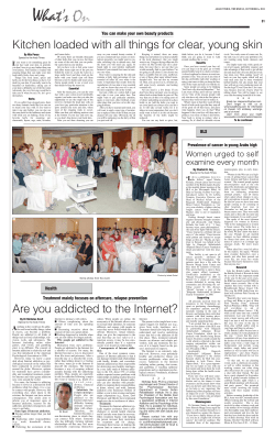


© Copyright 2025