
Tech. Sheet
Department of
Clinical Physiology,
Nuclear Medicine & PET
Annual Report 2011
Rigshospitalet · University of Copenhagen
Rigshospitalet
King Frederik V founded Rigshospitalet in 1757. Today, it has 1,200 beds, 8,500 employees
and an annual budget of 5 billion DKK. Research at Rigshospitalet is published in more
than 2,000 peer review papers per year, including around 90 higher academic theses (PhD
and Doctor of Medical Science). Rigshospitalet is part of The Capital Region of Denmark
and is a Copenhagen University Hospital.
University Of Copenhagen
The University of Copenhagen was founded in 1479. The Faculty of Health Sciences has
4,000 students and 10 bachelor- and master educations, including medicine and bioengineering in collaboration with The Technical University of Denmark, DTU. The University
of Copenhagen is member of IARU, The International Alliance of Research Universities.
www.ku.dk
Contents
Preface.......................................................................................................................................... 2
Mission and Objectives.............................................................................................................. 4
Organisation and staff................................................................................................................ 6
Highlights 2011......................................................................................................................... 10
Opening of the new PET/MR scanner 2011 ......................................................................... 12
Radionuclide therapy............................................................................................................... 14
Nuclear medicine 2011............................................................................................................. 16
Pediatric nuclear medicine...................................................................................................... 18
Cyclotron Unit........................................................................................................................... 20
Radiochemistry......................................................................................................................... 22
PET/CT scanning in oncology................................................................................................ 24
PET/CT scanning in radiation therapy................................................................................. 26
Cardiac PET .............................................................................................................................. 28
PET scanning of the brain........................................................................................................ 30
Collaboration with Landssygehuset, Faroe Islands............................................................ 32
Greenland - Iceland.................................................................................................................. 33
Academic activities................................................................................................................... 34
Patient investigations 2011...................................................................................................... 36
Finance....................................................................................................................................... 38
Publications 2011...................................................................................................................... 40
Research..................................................................................................................................... 48
Cluster for Molecular Imaging................................................................................................ 52
Danish Chinese scientific collaborations............................................................................... 55
Ib Henriksen Award 2011......................................................................................................... 56
Pasteur Prize 2011..................................................................................................................... 57
Young investigator prizes........................................................................................................ 58
The XV Scandinavian Congress of Clinical Physiology & Nuclear Medicine..................... 59
CIMBI Center for Integrated Molecular Brain Imaging....................................................... 60
Education................................................................................................................................... 62
Nuclear medicine technologists.............................................................................................. 64
PET and PET hybrid scanners ................................................................................................ 70
Equipment 2011......................................................................................................................... 73
European Medical Research Councils................................................................................... 74
Accreditation............................................................................................................................. 76
Preface
Liselotte Højgaard and Linda M. Kragh
Department of Clinical Physiology, Nuclear Medicine & PET was graced by a generous
donation from The John & Birthe Meyer Foundation for a new simultaneous whole body
PET/MRI scanner. It was opened at a Grand Occasion with an international symposium
”The John & Birthe Meyer PET Symposium in Copenhagen” 14th of December 2011. The
generous donation of 35 mio DKK was highly acknowledged by the department and the
hospital on the occasion, graced by the presence of Birthe Meyer, Gitte Meyer Brandt and
Søren Drost-Nissen from the foundation. You can read more about the celebration on
page 12. We are immensely grateful to The John & Birthe Meyer Foundation.
Our year 2011 was a busy year with 52,275 patient investigations, a significant rise compared to the year previously. The research effort resulted in 133 publications, 5 theses
(Ph.D. and Doctor of Medical Science) and 1 patent. We had a tough January 2011 with a
dramatic budget cut. Thank you to every one in the department for an amazing tenacity
and hard work to overcome the difficult beginning of the year.
It is the aim of our department that our patients should receive the best and most correct
patient investigation without delay for each and every patient. To reach our goals we
need to focus on research, innovation, education and quality.
We would like to convey our most sincere thank you to all staff members for making it
possible. Our aim is also to be a nice place to work. In line with that we were one of the
2
Department of Clinical Physiology, Nuclear Medicine & PET
best departments in the year 2011 qustionaire “TrivselOP” describing how the staff perceived their daily work. We were also among the best departments in the hospital in the
evaluation of our leadership competences. Thank you to every one for the great effort.
Apart from the grand opening of the PET/MR we got a new 64 slice PET/CT “mCT” from
Siemens Spring 2011 and we now have 4 PET/CT scanners in the department and the brain
PET research tomograph HRRT+.
In Nuclear Medicine the treatment with 177Lu-Dotatate in patients with neuroendocrine
tumors continued to grow with now more than 100 treatments per year. Clinical Physiology, Nuclear Medicine & PET is a Global Excellence department and the neuroendocrine
tumor program is accreditated by The European Neuroendocrine Tumor Society (eNETS).
A warm thank you to Professor Andreas Kjær who is head of the research in the department
and a warm thank you to all the researchers and the staff teaming up to make research the
basis for the best patient diagnosis and treatment throughout all activities in the department.
A warm thank you to Professor Gitte Moos Knudsen and her team at CIMBI for the enthusiastic and productive collaboration in neurobiology and research. (www.cimbi.dk)
A warm thank you to Head of Clinical Engineering Ole Bergsten for helping us with
equipment.
A big thank you to our directors at Center of Diagnostics, Rigshospitalet, Center Director, Dr. Bettina Lundgren and Leading Chief Technologist Karin Nørgaard and their
team for positive and helpful collaboration.
Our department has kept the budget now for the last 12 years. We acknowledge the needed
budget cuts, it was however a challenge to deliver a rising production of 10% for both patient investigations and R & D and at the same time implement the significant budget cuts.
Thank you to the Board of Directors, Rigshospitalet, where Torben Stentoft, Chief Executive Oficer, Jannik Hilsted, Chief Medical Officer and Helen Bernt Andersen, Chief Nursing Officer have been helpful in many ways throughout the year.
Thank you to the Department of Radiation Therapy for patience throughout the installation of the PET/MR scanner and for the daily great collaboration on research and patient
treatment. A special warm thank you to Professor Svend Aage Engelholm, Chief Nurse
Kirsten Amsinck and Chief Physicist Jens Peter Bangsgaard.
Internationally the EMRC, the European Medical Research Councils, with the office at ESF
in Strasbourg are thanked warmly for the great effort for strengthening medical research
in Europe and globally as seen on the last page of this report. Thank you to Dr. Stephane
Berghmans, Dr. Kirsten Steinhausen and Mrs. Janet Latzel and the whole team at EMRC.
Thank you to all staff members and collaborators in Denmark and internationally. Our aim
is to deliver the best patient treatment and the best research and education. Without your
great help we would not have made it.
Liselotte Højgaard Professor, Head of Department Linda M. Kragh
Chief Techonologist
Annual Report 2011
3
Mission and objectives
The mission of Rigshospitalet is to be the leading hospital in Denmark for patients in
need of highly specialized treatment.
The general objectives are:
» to be at the forefront of highly specialized diagnostic treatment and nursing
» to carry out research and development at an advanced international level
» to educate staff in the health services to a highly specialized level
» to contribute with professional advice and exchange of knowledge and expertise to
the wider healthcare community
» to be characterized by openness and human respect
The objectives of the Department of Clinical Physiology, Nuclear Medicine & PET are:
» to provide optimal clinical physiology and nuclear medicine for patient investigation
» to carry out research at the highest international level in clinical physiology and
nuclear medicine with special emphasis on molecular imaging, isotopes and
radiopharmaceuticals
» to deliver undergraduate and postgraduate education for all relevant professionals
within the relevant expert clinical fields, nationally and internationally
» to provide a good patient experience and ensure the wellbeing of the staff
The staff have participated in very many congresses, symposias, meetings and
workshops with invited lectures, oral presentations, abstracts and posters. We have
a comprehensive program for all staff members at the department, and frequent
visits from Danish and international research groups.
In 2011 more than 300 groups and individuals visited the department.
4
Department of Clinical Physiology, Nuclear Medicine & PET
Annual Report 2011
5
Organisation and staff
Physicians
Benzon, Eric von, MD, Chief Physician., Berthelsen, Anne Kiil, MD, Chief Physician., Borgwardt, Lise,
MD, PhD, Chief Physician., Christensen, Charlotte Birk, MD, Senior Registrar., Costa, Junia, MD,
Senior Registrar., Fischer, Barbara Malene, MD, Registrar, Ghotbi, Adam Ali, MD, Registrar., Hansen,
Tine Willum, MD, Senior Registrar., Hasbak, Philip, MD, Chief Physician., Hesse, Birger, MD, DMSc,
Chief Physician., Højgaard, Liselotte, MD, DMSc, Head of Department, Professor., Høyer, Alice Outzen
Widding, MD, Consultant., Jakobsen, Annika Loft, MD, PhD, Chief Physician., Jørgensen, Simon Møller,
MD, Registrar., Kjær, Andreas, MD, DMSc, PhD, MBA, Chief Physician, Professor., Korsholm, Kirsten, MD,
Registrar., Law, Ian, MD, PhD, Chief Physician., Löfgren, Johan, MD, Chief Physician., Markova, Elena,
MD, Chief Physician., Marner, Lisbeth, MD, Registrar., Mladenovic, Maja, MD, Registrar., Mortensen,
Jann, MD, DMSc, Chief Physician, Ass. Professor., Oturai, Peter, MD, Chief Physician., Özdemir, Zehra,
MD, Registrar., Rønne, Frederik, MD, Registrar., Winkler, Christine, MD, Senior Registrar.
PhD students, postdocs & clinical assistants
Andersen, Julie Bjerglund, MD, PhD Student., Andersen, Valdemar Lykke, Pharmacist, PhD Student,
Cimbi Researcher, Binderup, Tina, MSc, PhD Human Biology, postdoc , Malene M. Clausen, MD, PhD
Student, da Cunha-Bang, Sophie MD, PhD Student, NRU Researcher, Ettrup, Anders, MSc Human
Biology, PhD, NRU Researcher, Fisher, Patrick, PhD, NRU Researcher, Frøkjær, Vibe, MD, PhD, NRU
Researcher, Grüner Julie Marie, MD, PhD Student., Hag, Anne Mette Fisker, MSc Human Biology, PhD
Student., Haahr, Mette, MD, PhD Student, NRU Researcher., Hansen, Hanne Demant, PhD Student, NRU
6
Department of Clinical Physiology, Nuclear Medicine & PET
Head of Department
Professor Liselotte Højgaard, MD, DMSc
Chief Nuclear Medicine Technologist
Linda M. Kragh
Professor, Chief Physician
Andreas Kjær, MD, DMSc, PhD
Chief Physicist Søren Holm
Chief Physicist
Thomas Levin Klausen
Head Medical Secretary
Vibeke Rønn
KF Section
Chief Physician
Jann Mortensen,
MD, DMSc
Deputy Chief NMT
Tim Lundby
Computer Scient.
Flemming Andersen
Whole Body
Counter
PET scanner
section
Chief NMT
Linda M. Kragh
Chief Physician
Annika Loft Jakobsen
MD, PhD
Chief Physician
Peter Oturai
Cyclotron Chief
Holger Jensen,
MSc, PhD
Deputy Chief NMT
Kate Pedersen
Cyclotron Unit
Cyclotron Chief
Holger Jensen,
MSc, PhD
Radiochemistry
Unit
Chief Radiochemist
Production Manager
Jacob Madsen
Chief Radiochemist
Nic. Gillings
Deputy Chief NMT
Anne Sørensen
Department of Clinical Physiology, Nuclear Medicine & PET is part of The Diagnostic Center headed by Bettina Lundgren,
Director, MD, DMSc and Karin Nørgaard, Vicedirector.
Researcher, Hansen, Martin, MSc, PhD Student., Herth, Matthias, PhD, Cimbi Researcher, Hollensen,
Christian, PhD Student., Jensen, Mette Munk, MSc Human Biology, Research Fellow., Johnbeck, Camilla
Bardram, MD, PhD Student., Jørgensen, Jesper Tranekær, MSc Human Biology, Research Fellow.,
Knudsen, Jesper Andreas, MD, PhD Student., Kristoffersen, Ulrik Sloth, MD, Research Fellow, PhD
Student., Li, Fan, MSc Med Tech, PhD Student., MacMahon, Brenda MD, PhD Student, NRU Researcher,
Marthin, June, MD, PhD Student., Nedergaard, Mette Kjølhede, MD, PhD Student, Nielsen, Anna Pors,
MD, Research Assistant, NRU Researcher, Nielsen, Carsten Haagen, MSc Med Tech, PhD Student.,
Nielsen, Kristina Rue, MD, PhD Student., Olesen, Oline Vinter, MSc Med Tech, PhD Student., Persson,
Morten, MSc Pharm, Research Fellow, PhD Student., Perdersen, Sune Folke, MSc Human Biology, PhD
Student., Pfeifer, Andreas, MD, Research Fellow, PhD Student., Reichkendler, Michala, MD, PhD Student.,
Ripa, Rasmus S, MD, DMSc, postdoc, Skovgaard, Dorthe, MD, PhD, Registrar., Zornhagen, Kamilla
Westarp, DVM, PhD Student.
Physicists, pharmacists, chemists, technicians, engineers, computer scientists, QA-assistants
Andersen, Flemming, MSc, PhD, Computer Scientist., Boudreault, Ghislain, MSc, PhD, Substitute
Cyclotron Physicist., Brandt-Larsen, Malene, MSc, PhD, Chemist., Christensen, Jan Damgaard, Cyclotron
Technician., Dahan, Daniel, Cyclotron Technician., Dähnhardt, Andreas, Computer Assistant., de Nijs,
Robin, Medical Physicist, MSc, PDEng, PhD., Denholt, Charlotte Lund, MSc, PhD, Chemist., Erlandsson,
Maria, Chemist., Gillings, Nicholas, MSc, PhD, Chief Radio Chemist., Heilmann, Helene, QAPharmacist., Holm, Søren, MSc, PhD, Chief Physicist., Jensen, Björn Neumann, Electro Engineer., Jensen,
Annual Report 2011
7
Holger, MSc, PhD, Physicist, Cyclotron Chief., Jensen, Tina Gade, QA-Assistant., Jørgensen, Jesper,
MSc, Cyclotron Physicist., Keller, Sune Høgild, MSc, PhD, Computer Scientist., Klausen, Thomas Levin,
MSc, Chief Physicist., Lehel, Szabolcs, MSc, PhD, Chemist., Madsen, Jacob, MSc, PhD, Chemist, Chief
Production Manager., Schjøth-Eskesen, Christina, Chemist., Sibomana, Merence, IT-expert., Weihrauch,
Per, Cyclotron Technician.
Nuclear medicine technologists (NMT), radiographers and nurses
Abrahamsson, Elisabeth, Radiographer., Albers, Mia C. Hjorth, NMT., Bojesen, Christina, NMT.,
Christensen, Camilla, NMT., Christensen, Pia, NMT., Cortsen, Annette, NMT., Dall, Bente, NMT., Dunbar,
Douglas, NMT., Elkington, Sakeena, NMT., Federspiel, Marianne, NMT., Frederiksen, Mette Borggreen,
NMT., Hansen, Anja Vallin, NMT., Hansen, Jon, Radiographer., Hansen, Lasse, NMT., Hassan, Mariam,
NMT., Heiberg, Therese, Cand.scient.san, NMT., Holm, Stine, NMT., Jensen, Charlotte, NMT., Jensen,
Martin Ravn, NMT., Jørgensen, Hanne, NMT., Jørgensen, Mette Møller, NMT., Kernchen, Ulla, Staff
Nurse., Knudsen, Camilla Sloth, NMT., Kragh, Linda M., Chief NMT., Kronvall, Johanna, NMT., Lindell,
Elin, NMT., Linnet, Solveig, NMT., Ljunggren, Anna, NMT., Lundby, Tim, Deputy Chief NMT., Lærke,
Sonja Pedersen, Laboratory Technician., Magnusson, Linda, Technologist., Myschetzky, Rebecca, NMT.,
Nehme, Ghina, NMT., Osinska, Dorota, NMT., Osmani, Lume, NMT., Pedersen, Kate, Deputy Chief NMT.,
Pejtersen, Maria, NMT., Poulsen, Camilla Storm, NMT., Saxtoft, Eunice, NMT., Setterberg, Victoria, NMT.,
Sørensen, Anne B., NMT., Sørensen, Anne, Deputy Chief NMT., Sørensen, Louise Sørup, NMT., Stahr,
Karin, NMT., Svalling, Susanne, NMT., Wikke, Tina, NMT., Yerst, Joo, NMT., Zulfovska, Cejlan, NMT.
8
Department of Clinical Physiology, Nuclear Medicine & PET
Medical secretaries and secretaries
Frydendall, Pia, Medical Secretary., Hansen, Lissa, Medical Secretary., Hildebrand, Sanne, Secretary.,
Marquardsen, Joan, Medical Secretary., Myltoft, Mette Gylling, Medical Secretary., Nielsen, Mariane,
Service Assistant., Nielsen, Tina Vikmann, Medical Secretary., Runge, Gitte, Medical Secretary., Rønn,
Vibeke, Head Medical Secretary., Semitoje, Gudrun, Medical Secretary., Stahlfest, Marianne, Medical
Secretary.
Students
Christensen, Anders Nymark., Bothmann, Bianca., Dohn, Asmus Ougaard., Dyring, Maria., Henning,
William Sebastian., Henriksen, Martin Romme., Husted, Frederik Agner., Ipsen, Niels Bruun., Jensen,
Simon Bøgh., Larsen, Thorvald Wadum., Lassen, Martin Lyngby., Nielsen, Anders Bo., Nørgaard, Martin.,
Øberg, Mikkel., Paamand, Rune., Perez, Nicolas Palm., Petersen, Stefan Alaric., Savic, Dragana., Værge,
Thorkil Kowalski., Weihrauch, Simone., Yildiran, Handan
Annual Report 2011
9
Highlights 2011
Søren Holm, Master of Science PhD and Responsible
Physicist at the department, celebrated his 25 years
anniversary at Rigshospitalet on 11th of November 2011
with a symposium with lectures in Auditorium 2 and a fine
reception followed the scientific program.
Consultant Birger Hesse, MD, DMSc, a world renowned
capacity in nuclear cardiology, with a long duty for the
speciality in Denmark and internationally, celebrated his
Retirement Symposium on 14th of January 2011. A large group
of invited guests and distinguised speakers made it a very
successful day. From the department we would like to express
our sincere gratitude to Birger Hesse for his long and important
duty for the strengthening of clinical physiology and nuclear
medicine, not only at our department here at Rigshospitalet,
but also in Denmark and internationally.
Oline Vinter Olesen, Bioengineer in Medicine and
Technology, defended her PhD thesis: “Markerless 3D Head
Tracking for Motion Correction in High Resolution PET
Brain Imaging” on the 15th of December 2011. Oline has
invented a new technology for motion correction in brain
PET, tested at Yale in the US and at Rigshospitalet and
developed in collaboration with Professor Rasmus Larsen,
DTU Informatics, Technical University of Denmark, Siemens
Healthcare and Rigshospitalet.
The department opened the new DEXA-scanner in 2011,
so that routine clinical patients from Rigshospitalet can be
refered directly to our department for investigations.
10
Department of Clinical Physiology, Nuclear Medicine & PET
The old PET/CT scannner from 2001,
one of the first in Europe, was outdated
and exchanged by a new PET/CT
scanner a Siemens MCT with 64 slice
CT. The department has now 4 PET/CT
scanners of state of the art quality.
A new Siemens Symbia 16
slice SPECT/CT
was opened November 2011.
The new advanced
FDG production lab
was opened in 2011.
The new textbook in clinical nuclear medicine was
published in 2011 edited by Dr Charlotte Birk
Christensen, Dr Annika Loft Jakobsen and Dr
Birger Hesse from our department and with many
contributions to chapters from the rest of the staff
Nuclear Medicine Technologist Susanne Svalling got the
”Arbejdsmiljøpris 2011” at the “Arbejdsmiljødag 2011”
on Rigshospitalet
Annual Report 2011
11
Opening of the new PET/MR
scanner 2011
- donated by the John and Birthe Meyer Foundation
The absolute Highlight of the year 2011 was the generous donation from the John and
Birthe Meyer Foundation of 35 mio DKK for a new integrated PET/MR whole body scanner. The opening of the new scanner took place 14th of December 2011 under the presence
of Birthe Meyer, Gitte Meyer Brandt and Søren Drost-Nissen from the foundation. It was
a day of celebration and joy and the gratefulness from the whole department and the
hospital was conveyed to the foundation. The gratitude from the whole community of researchers towards Birthe Meyer, Gitte Meyer Brandt and Søren Drost-Nissen and the John
and Birthe Meyer Foundation was expressed very clearly from every one.
In the morning we had an international symposium on the use of PET and MR for research and clinical diagnosis, with our new collaborator Professor Bruce Rosen from the
Martinos Center, Harvard Medical School. Bruce Rosen received the prestigous K.F.J.
Award the next day, as a recognition of his cutting edge research in neuroimaging. Professor Gitte Moos Knudsen from CIMBI, Rigshospitalet is Visiting Professor there to
establish and strengthen the collaboration between our two institutions. Both Gitte and
Bruce gave inspiring talks about the future use of PET/MRI in the neurobiology research
program. Professor Søren Bentzen from University of Madison Wisconsin is an expert on
PET and MR in radiation therapy planning and medical physics and also adjunct profes12
Department of Clinical Physiology, Nuclear Medicine & PET
sor here at the University of Copenhagen. He gave his vision for the use of PET/MR for
radiation therapy planning in the future and stressed the importance of interdisciplinarity. Head of Department of Radiotherapy Professor Svend Aage Engelholm reported on
the very successful synergy between radiotherapy and PET research in Copenhagen,
where we have been game changers for the use of PET/CT planned radiotherapy. At present we perform 1000 clinical patients with PET/CT planning. Professor Lena Specht is a
world authority on the use of PET/CT for radiation therapy planning in head and neck
cancer and malignant lymphoma. She underlined the significance of PET/CT and PET/
MRI for improved therapy in the future. Professor Andreas Kjær gave a state-of-the-art
perspective on the use of molecular imaging PET/MR as a very important translational
research tool. Dr. Ian Law reported on the future use of PET/MR for brain tumors and dementia. Dr. Annika Loft Jakobsen, Head of our PET section, and Dr. Anne Kiil Berthelsen
gave an elegant presentation on the use of PET/CT and future use of PET/MRI for oncology in combined research and clinical investigations. Finally Dr. Lise Borgwardt reported
on the use of PET/CT and future use of PET/MR in pediatric nuclear medicine. Head of
Department Professor Liselotte Højgaard summarized the history of PET in Copenhagen:
The first PET center in Denmark and the first of the Nordic countries with PET/CT and
now PET/MRI.
Annual Report 2011
13
Radionuclide therapy
131
I-Radioiodine and 177Lu-DOTATATE
Peter Oturai and Jann Mortensen
For many years the department has successfully treated patients with benign thyroid
diseases – goiter and hyperthyroidism – with 131I-iodine.
Since May 2009 we have provided treatment with 177Lu-DOTATATE in Denmark to
patients with neuroendocrine tumor (NET) metastases. Until the end of 2011 58 patients
have been given a total of 187 treatments. Being one of the two highly specialized centers using this treatment modality in Denmark we work in close collaboration with the
Departments of Gastro Surgery, Oncology, Radiology, Pathology and Endocrinology at
Rigshospitalet.
The NET-Center at Rigshospitalet has been accredited as Center of Excellence by the
European Neuroendocrine Tumor Society (eNETS).
Lu-DOTATATE is synthesized and labelled at the Hevesy Laboratory at Risø, DTU.
Lu-DOTATATE is administered in our dedicated facilities in the department and the
patients stay overnight at the surgical ward.
177
177
The rationale for the treatment, is that the radio labelled somatostatin analogue binds
to neuroendocrine tumors expressing somatostatin receptors. The emitted beta-particles
from the 177Lu-isotope destroy the tumor cells. In addition the emission of gamma photons from 177Lu allows for scintigraphic imaging and dosimetry.
14
Department of Clinical Physiology, Nuclear Medicine & PET
The indications for 177Lu-DOTATATE therapy are inoperable patients with neuroendocrine tumors that either show progression or cannot tolerate standard treatment. A
prerequisite is an 111In-octreotide scan demonstrating a high density of somatostatin
receptors in the tumors.
Our experience is comparable to what we expect from the literature, i.e. some measurable effect in the majority of the patients, and only few side effects.
Annual Report 2011
15
Nuclear medicine 2011
Jann Mortensen and Peter Oturai
The majority of our more than 30,000 patient examinations in the department are related to the diagnosis and monitoring of cancer patients. Hybrid SPECT/CT scans of
neuroendocrine tumors, bone scans for primary and secondary malignant tumors, sentinel node scintigraphy for breast cancer, malignant melanoma and other malignancies,
MUGA and 51Cr-EDTA clearance measurements for monitoring side effects in chemotherapy treated patients are some examples.
The somatostatin receptor ligand 111In-Octreotide imaging, most often combined with
CT scans of diagnostic quality, is an important endocrine nuclear medicine imaging
modality and is being increasingly used for evaluation and monitoring of therapy in
patients with inoperable tumors.
Frequent indications for lung physiology measurements are control after chemotherapy
and transplantation or pre- and postoperative evaluation and radioaerosol mucociliary
clearance investigations for the diagnosis of primary ciliary dyskinesia.
Radioisotope leakage monitoring procedures are used during isolated limb perfusion
with melphalan and tumor necrosis alpha for recurrent melanoma and soft-tissue sarcoma. To this end, we have eight gamma cameras for routine nuclear medicine imaging
and research studies, three hybrid SPECT/CT cameras, one dual-head gamma camera
and four single-head cameras. A new SPECT-16 slice-CT (Symbia, Siemens) was in16
Department of Clinical Physiology, Nuclear Medicine & PET
stalled and replaced our Hawkeye hybridscanner. This new scanner will primarily be
dedicated to the increasing number of pediatric nuclear medicine investigations in the
department.
We use our ultrasound scanner for thyroid patients as a supplement to thyroid scintigraphy. For lung function testing we have two Jaeger body plethysmographs. At the
end of the year we had our first DEXA scanner for bone mineral content and body
composition assessment installed. Our animal facilities are equipped with SPECT and
PET and CT scanners. Weekly receptor targeted radionuclide therapy against neuroendocrine tumors, initiated 2009, is now a routine function in our department. Read more
about 177Lu-DOTATATE therapy on page 14.
In 2011 we have had weekly and monthly conferences dealing with thyroid diseases,
neuroendocrine tumors, pediatric oncology, pulmonary, cardiology, adrenal and orthodontic diseases.
Annual Report 2011
17
Pediatric nuclear medicine
Lise Borgwardt
Each year we perform 1500 pediatric nuclear medicine investigations, thereof 300 PET
scans, mainly for the large pediatric clinics at the hospital. It is a special focus area for our
department to perform these investigations at the highest level of excellence, and at the
same time make it a positive experience for both the child and its parents. The department
is a member of the EANM Pediatric Committee, Pediatric Nuclear Medicine Network and
the International Telemedicine Network for Second Opinion and Exchange of Ideas.
Our Pediatric Focus Group is still evolving. Education internally in the department, to the
referring departments at the hospital, and also externally, is a high priority. This is why
our Pediatric Focus Group this year contributed at the NML congress in Copenhagen and
gave several talks on different angels of pediatric nuclear medicine with great success.
Also as invited speaker at IPR, International Pediatric Radiology 2011, the group gave a
talk on future perspectives on hybrid scanners in Pediatric Nuclear Medicine. The group
contributed to the Danish book on Clinical Nuclear Medicine, naturally with the chapter
on Pediatric Nuclear Medicine. Also the work in the Pediatric Committee has been very
interesting and we will continue this process.
We have been performing PET/CT for radiotherapy planning in children since 2003, performed by our pediatric radiotherapy-planning-team Anne Kiil Berthelsen, Annika Loft Jakobsen, Charlotte Birk Christensen and Morten Jørgensen. We have now performed more
than 60 PET/CT for radiotherapy planning in children. This year we received the prize for
18
Department of Clinical Physiology, Nuclear Medicine & PET
the most important scientific work at the ESOPNM, European Society of Pediatric Nuclear
Medicine, 2011 for our survey on our experience with PET/CT for radiotherapy planning
in children. Together with the Department for Radiotherapy at Rigshospitalet and our new
collaboraters across the Øresund strait; Thomas Björk-Eriksson, Section for Radiotherapy,
Skåne University Hospital in Sweden, the group received 450,000 DKK in fundings from
the Danish Children Cancer Foundation to further develop this important area.
Diagnostics on children with Hodgkin’s lymphoma follows the guidelines of the European
protocol and we participate in the teleconferences in the European Pediatric Hodgkin’s
Network Group to increase the level of interpretation in Europe in general. On all children
for PET scanning, including the children with lymphoma, we perform a heating procedure
without medication in order to diminish uptake in activated fat, with great results.
Research in Pediatric Nuclear Medicine and PET is necessary, as we have an increasing
amount of medical doctors, students and technicians involved in the field and we conduct research protocols in children with PET/CT in lymphomas, sarcomas, metabolismand perfusion PET in neonatals, diagnostic value of HIDA-scintigraphy and the optimal
preparation of the child with suspicion of biliary atresia, MIBG SPECT/CT and PET/CT
of children with neuroblastomas in coorporation with Ludwig Maximilian University
of Munich etc. In the beginning of the year we were visiting Chief Physician Thomas
Pfluger, Ludwig Maximilian University of Munich for further collaboration in between
our institutions.
In 2011 we received a new SPECT/CT scanner dedicated for the children. We have been
performing highend hybrid SPECT/CT in children since 2003 and we look forward to
develop this strategy even further with the new opportunity.
We are futhermore expecting an interesting year 2012 with the new PET/MRI scanners
arrival in December this year and the very important possiblities for new studies in children on this new hybrid modality.
Annual Report 2011
19
Cyclotron Unit
Holger J. Jensen
In 2011 we experienced, as in the previous years, an increase in our production of radioactive isotopes and ended up with approximately 1148 successful productions (602
and 546 productions for our Scanditronix MC32 and Siemens RDS Eclipse cyclotrons
respectively) or 16.8% more than in 2010. During the last 6 years we have seen in
average an increase of 6% more productions per year. The major production increase
this year came from 11C, which increased with 68.4%, whereas our production of 18F
only increased by 1.4%. All other isotopes stayed at a constant level. The year was
relatively quiet without any major technical problems for the two cyclotrons and due
to the high flexibility of running two cyclotrons we succeeded to have less than 1%
cancelled productions in 2011.
Looking at the development in the produced activity, we have increased the activity by
16% per year in average since 2006. Despite of this we have managed to keep the doses
to the employees in the cyclotron- and radiochemistry unit under control and managed
to reduce the doses during the last few years as seen in figure 2.
Also for the release of radioactive gas waste from our production, we have seen a
positive development. As seen in figure 3, we have managed to reduce the release
despite of the increased production.
20
Department of Clinical Physiology, Nuclear Medicine & PET
Figure 1. Development in productions since 2006
1400
Productions
1200
1000
800
600
400
Total
200
18F
0
11C
2006
2007
2008
2009
2010
2011
Figur 3. Release of radioactive gas waste from our
production since 2006
2,5
3000
2,0
2500
Release [GBq]
Dose [mSv]
Figure 2. Average received doses for employee in the
cyclotron and radiochemistry group since 2006
1,5
1,0
0,5
0,0
2006
2007
2008
2009
2010
2011
2000
1500
1000
500
0
2006
2007
2008
2009
2010
2011
Annual Report 2011
21
Radiochemistry
Nic Gillings and Jacob Madsen
2011 has been a very productive year for the radiochemistry section. Overall productivity
has increased considerably compared with previous years (see figures below) with a notable increase in productions for animal and in vitro studies (preclinical research). Three
new PET radiopharmaceuticals were approved for human use in 2011, with a fourth
expected in early 2012, which will bring the total up to 14. Implementation of several
other new radiopharmaceuticals for human use is underway. Permanent staff currently
consists of 4 medical laboratory technologists, 1 pharmacist (QA) and 5 radiochemists. In
addition there are 2 post-docs and 3 Ph.D. students working in radiochemistry.
Routine production
The demand for 18F-FDG increased slightly in 2011, with the total amount of radioactivity
produced increasing by approximately 10%. A new FDG production laboratory has been
built, validated and approved by the Danish Medicines Agency. Routine production is
gradually being transferred to this laboratory in order free up space for production of
new radiopharmaceuticals in the existing laboratory. 18F-Fluoroethyl tyrosine (18F-FET)
is now produced on a weekly basis for clinical brain scans of up to 16 patients from each
batch. Production of krypton-81m generators continued according to the well-established
delivery schedule on Mondays, Wednesdays and Fridays.
Research production
2011 was a busy year, with regular production of a large range of radiopharmaceuticals
22
Department of Clinical Physiology, Nuclear Medicine & PET
for research use, both for humans, animals and in vitro studies. Productions for human
neuroreceptor imaging increased dramatically in 2011 (up approx. 80%) and the number of animal studies also increased. Production of the Alzheimer tracer, 11C-PIB continued
at a similar level as in 2010. Production of radiopharmaceuticals for oncology research
continues to increase. The cell proliferation tracer, 18F-FLT, is now being produced routinely
for clinical research studies and production of the neuroendocrine tumor tracer,
68
Ga-DOTATOC, for human use will commence in early 2012. Animal studies are ongoing
using 64Cu-ATSM (hypoxia tracer), 18F-Annexin V (apoptosis tracer), 64Cu-DOTA-AE105
(urokinase-type plasminogen activator receptor (uPAR) tracer) and radiolabelled RDG
peptides (angiogenesis tracers).
Radiopharmaceutical development
Collaboration with The Neurobiology Research Unit, Rigshospitalet and the Department of Medicinal Chemistry at PHARMA (University of Copenhagen) under CIMBI
(Centre for Integrated Molecular Brain Imaging) continued in 2011. Production of
11
C-Cimbi-36, the very promising 5-HT2A agonist PET tracer developed over the last
few years, has been fully validated and we hope to get permission for human studies
in early 2012. On the new development front, work is in progress on development of
a 5-HT7 PET ligand and a fluorine-18 analogue of 11C-Cimbi-36. Initial results are encouraging and this work will continue in 2012. In 2011 a new Ph.D. student started to
continue our program on development of labelled peptides for imaging the epidermal
growth factor tyrosine kinase receptor (EGFR). This research is performed in collaboration with The Department of Natural Sciences, University of Copenhagen. Synthesis of
the activated ester 18FSFB has been optimized, fully automated and used in the synthesis of 18F-Annexin V. After initial challenges with the protein purification 18F-Annexin V
is now regularly synthesized in very high radiochemical purity. Another ongoing project involves development of a new tracer for uPAR imaging. Beside 64Cu-DOTA-AE105
(mentioned above) alternative labelling strategies for the introduction of Galium-68 or
Fluorine-18 are being pursued.
900
800
700
600
500
400
300
200
100
0
Radiopharmaceutical productions for animal and
in vitro studies
120
No. of productions
No. of productions
Radiopharmaceutical productions for human use
2006
2007
2008
2009
2010
2011
100
80
60
40
20
0
2006
2007
2008
Nuclear Medicine & PET
Neurobiology Research Unit
[81mKr]Krypton generators
2009
2010
2011
Year
Year
PET & Cluster for Molecular Imaging
Neurobiology Research Unit
[18F]FDG
Annual Report 2011
23
PET/CT scanning in oncology
Positron emission tomography is now well established in oncology and plays a major role
in the diagnostic work-up for many patients due to the high sensitivity and specificity for
diagnosis and treatment response monitoring.
Our CT scans of PET/CT are performed as high quality diagnostic scans with the use of
oral and intravenous contrast media. The PET- and the CT scans are initially interpreted
separately followed by a joint interpretation of the fused images and a final, combined
conclusion taking both examinations into account. This provides the clinician with a more
precise PET result, a better CT result, and also a more useful conclusion. The CT result
improves in quality because PET can help depicting small tumors that could easily have
been overlooked even by a trained radiologist’s eye. The PET positive foci are more precisely determined as correct or false positive with the help of the CT information. Finally,
the combined PET/CT conclusion is superior to both scan results alone. Furthermore, the
patient is spared from an extra CT examination at the Department of Radiology as well as
an extra radiation dose.
In 2011 we performed 6000 PET/CT scans, and more than 90% were oncological. The
indications are mainly diagnosis, staging, therapy monitoring and detection of recurrent
disease in patients with a variety of malignant diagnoses. As a routine, we have included
a supplementary CT of the lungs with breathhold tecnique to improve the diagnostic
quality. Approximately 50% of our patients participate in clinical research protocols. Our
24
Department of Clinical Physiology, Nuclear Medicine & PET
Annika Loft Jakobsen and Anne Kiil Berthelsen
main topics are gynecology, malignant lymphoma, neuroendocrine tumors and lung
cancer. We have just completed a study investigating patients with metastases from an
unknown primary. We are also studying patients with colorectal cancer in collaboration
with Næstved Hospital as well as we have an ongoing multicentre-study with Odense
and Aalborg University Hospitals investigating endometrial cancer.
We have 17 weekly multidisciplinary team conferences, where our PET/CT scan results
are discussed with the clinical experts.
FDG is still the main tracer in oncology, but we also use 18F-NaF, 18F-FET and 18F-FLT in
clinical studies as well as in research protocols.
Working with PET/CT for many years now, we are convinced of the usefulness of PET/
CT in the every day clinical work. However, clinical trials are still necessary to verify the
usefulness of the method, refine the scanning protocols and to develope new indications.
Annual Report 2011
25
PET/CT scanning in radiation
therapy
Annika Loft Jakobsen and Anne Kiil Berthelsen
The use of PET/CT in radiotherapy planning of cancer patients has increased very rapidly since
the method was introduced in 2001. The introduction of more advanced treatment methods,
e.g. RapidArc and IMRT, the precise tumor delineation, has become even more critical.
We cooperate closely with the Department of Radiotherapy on the use of PET/CT for
treatment planning of cancer and we perform 1000 PET/CT scans for the Department of
Radiotherapy every year.
The demanding collaboration between mould technicians, nuclear medicine physicians
and technologists, radiologists and radiology technologists, radiation oncologists, physicists, and dosimetrists must be emphasized.
The advantages are numerous: the anatomical localisation and the metabolic activity of
the tumor are defined, especially when the tumor density in CT images is difficult to differentiate from that of the surrounding normal tissue, and the tissue heterogeneity can then
be taken into account when choosing radiation technique and energy, and only one scan is
necessary. All our PET/CT scanners have the possibility of performing PET/CT scans for
radiotherapy planning. The nuclear medicine specialist delineates the viable tumors depicted by PET on the fused PET/CT images after interpretation together with the radiologist.
We rely on visual anlysis more than fixed threshold levels. The scans are always performed
as whole-body examinations that give us the opportunity to depict unknown metastatic
26
Department of Clinical Physiology, Nuclear Medicine & PET
disease. We have a 19% detection rate of metastases or new primary malignancies leading
to change of treatment. The regions are exported to the radiation dose planning system
together with the CT scan, and the information is incorporated in the treatment planning.
Research in this field is necessary, and we have conducted trials with nasopharyngealand cervical cancer and malignant lymphoma with encouraging results. PET/CT for radiotherapy planning is now used routinely for patients with brain, cervix-, head & neck-,
lung-, oesophageal-, cardia-, rectum- and anal cancer as well as malignant lymphoma and
mesothelioma. We have an ongoing study using breathhold PET in lymphoma of the mediastinum and we are collaborating in a multicentre study for lung cancer. We are looking
into recurrent head and neck cancer related to the primary PET tumor delineation.
A new research area is PET/CT for radiotherapy for children. We have used PET/CT for
radiotherapy planning for children for seven years and we are currently reviewing our
data upon this issue. Together with the radiation oncologists we are concerned about the
risk of developing secondary malignancies due to radiotherapy and we want to look into
the possibilities of reducing the radiation fields and introducing dose painting by PET.
We strongly believe that PET/CT based radiotherapy planning will improve the therapeutic output in terms of target definition and non-target avoidance and will play an important role in future therapeutic interventions in many malignant diseases.
Annual Report 2011
27
Cardiac PET
Philip Hasbak
Non-invasive assessment of myocardial function is an important domain of PET. Traditionally,
cardiac PET images have been visually interpreted, using uptake as a measure of function.
This approach, however, takes only a fraction of the full PET information into account. In
contrast, the application of tracer kinetic modeling to dynamically measured data is able to
extract objective measures of perfusion and/or metabolism, depending on the tracer. While
such true cardiac quantification has been troublesome and time-consuming in the past, our
new cardiac tool now makes this state-of-the-art technology readily available.
Research-wise we have a close and good cooperation with various clinical departments.
Certain ongoing projects should be emphasized:
In collaboration with
» Department of Cardiology: Molecular imaging in patients suspected of acute
ST-elevation myocardial infarction (STEMI) with angiographically normal coronary arteries.
» Department of Oncology and Radiation Therapy: Cardiac side effects in radiation
therapy for breast cancer
» Department of Vascular Surgery and Department of Anestesiology: Buttock ischemia
after endovascular abdominal aortic aneurysm repair
Cardiac SPECT
At Rigshospitalet there is a special need to provide cardiac SPECT every day, since a
28
Department of Clinical Physiology, Nuclear Medicine & PET
How to differentiate acute myocardial infarction (MI) from takotsubo cardiomyopathy with
quantitative cardiac PET
Quantification of myocardial blood flow in the acute
phase of MI. Zero-flow in the MI-area. Reduced to
normal flow in the tissue surrounding the MI (area at
risk?) and normal to increased flow in the basal part
of the left ventricle.
Quantification of myocardial blood flow in the very
acute phase of takotsubo cardiomyopathy revealed
normal flow between 0.78-1.07 ml/min/g (normal range
0.6-1.2 ml/min/g) in the apical bllooning part of the LV,
whereas the perfusion in the basis was increased.
large proportion of our patients with ischemic heart disease need an acute or subacute
work-up that requires quick decision making as to coronary revascularization strategy.
Patients with unstable angina or non-STEMI should be revascularized with percutaneous coronary intervention (PCI) within 3 days after admission or with coronary artery
bypass graft surgery (CABG) within 5-7 days according to The Danish National Board
of Health.
We are participating in the following trials: CORE320 (Brigham and Women’s Hospital),
CAMARO (Mayo Clinic), PRECISE (Cardiovascular Core Lab) and ST-segment study
(Cardiovascular Core Lab) and KAI (Cleveland Clinic)
Cardiac 123I-MIBG
In 2010 we introduced cardiac 123I-metaiodobenzylguanidine (123I-MIBG). Radiotracer
analogs of the sympathetic mediator norepinephrine have been investigated extensively,
and are at the brink of potential widespread clinical use, especially after the presentation
of the ADVANCE-HF trial. The most widely studied SPECT tracer, 123I-MIBG has consistently shown a strong, independent ability to risk stratify patients with advanced congestive heart failure. One ongoing project should be emphasized:
In collaboration with Department of Cardiology: Cardiac sympathetic activity in terminal
heart failure patients with and without ”left ventricular assist device” (LVAD)
Annual Report 2011
29
PET scanning of the brain
Ian Law
In 2011 the number of clinical PET and PET/CT scans grew even further than in 2010. The
investigations cover the range from neurooncology, neurodegenerative disease, cerebrovascular disease and epilepsy.
In the autumn for 2011 we introduced, as the first site in Denmark, an exciting new and
advanced PET tracer, 18F-Flouro-Ethyl-Tyrosine (FET), in neurooncology. FET is an aminoacid analogue that is preferably targeting primary brain tumors and is particularly helpful in tumor delineation. The intended clinical use is in treatment planning presurgically
and in radiation therapy, in tumor grading, in monitoring treatment effects and in differentiating viable tumor tissue and treatment damage. FET is ideal for large radiotracer
production, and we have felt a lot of interest from the clinicians.
Rigshospitalet is treating and monitoring all brain tumors on Seeland. Thus, the implementation of this unique radiotracer is an important milestone in our efforts to secure the
best clinical diagnostic methods available for our patients. Further, we have secured sufficient production capacity to accommodate the growing need in the future.
Continued development and research is one of our fundamental goals, and we will be
conducting studies focusing on the optimized use of PET FET in the primary radiotherapy
planning and in reirradiation, in surgical planning, in tumor grading, in pediatric brain
tumors and in the use of PET/MRI.
30
Department of Clinical Physiology, Nuclear Medicine & PET
Fig 1. Two patients referred for the differentiation of recurrent high grade tumor or treatment damage? The top
patient has recurrent and metabolically active tumor tissue (red arrow) and the bottom patient has treatment
damage (green arrow). The images shown are T1 weight MRI after contrast, PET FET scanning and FET PET
fused to MRI.
Fig 2. Patient with malignant high grade tumor treated with surgery, radiation-chemo-therapy and antiangiogenic
treatement (Bevacizumab). There is a large recurrent metabolically active tumor (red arrow) that is only partly
manifest on MRI. Other changes on MRI are not recurrent tumor (green arrow).
Annual Report 2011
31
Collaboration with
Landssygehuset, Faroe Islands
The Department of Clinical Physiology, Nuclear Medicine & PET, Rigshospitalet has a
close collaboration with Landssygehuset in Tórshavn, Faroe Islands.
The hospital has a staff of 850 (the largest employer in Faroe Islands) and 180 beds. The
9,000 in-patients and 35,000 out-patients annually are taken care of by 29 specialities, of
which 9 are via consultant collaborations, including collaboration in clinical physiology
and nuclear medicine with Rigshospitalet.
In 2011 the Department of Clinical Physiology and Nuclear Medicine in Tórshavn performed more than 500 scintigraphies of lungs, bones, thyroid, kidneys, sentinel nodes and
renography. The department has one 2-headed Skylight camera, a Norland DXA scanner and a Jaeger whole body plethysmograph. Second opinion on scintigraphy and lung
function measurements is provided via a direct telemedicine connection. The responsible
physician and phycisist for Nuclear Medicine in Tórshavn is Consultant, DMSc Jann
Mortensen and Physicist Thomas Levin Klausen.
32
Department of Clinical Physiology, Nuclear Medicine & PET
Greenland - Iceland
Jann Mortensen
and Thomas Levin Klausen
Rigshospitalet receives the patients from Greenland who need highly specialized
treatment. In our PET Center we also investigate patients from Iceland.
Annual Report 2011
33
Academic activities
Andreas Kjær, Professor, Chief Physician, is the
President of the Scandinavian Society of Clinical
Physiology and Nuclear Medicine (SSCPNM),
member of the Scientific Committee of the Danish Cancer Society, Editor-in-Chief of Open
Neuroendocrinology Journal as well as for Diagnostics. Leader of the project Molecular Imaging
for Testing of New Drugs funded by the Danish
National Advanced Technology Foundation,
Leader of a project on new PET tracers funded
by the Strategic Research Council and Partner
of the Danish Chinese Center for Proteases and
Cancer funded by the National Natural Science
Foundation of China and the Danish National
Research Foundation, National Center Director
and partner of EATRIS (the European Advanced
Translational Research Infrastructure in Medicine) under the EU 7FP, and Head of the Cluster
for Molecular Imaging and Director of the Postgraduate School for Medical & Molecular Imaging at the Faculty of Health Sciences, University
of Copenhagen.
Anne Kiil Berthelsen, Chief Physician is a
member of the ILROG, EORTC Lymphoma
Group, the Danish Radiology Society, the Danish
and the Nordic Society of Gynaecological Oncology, the Danish Society of Clinical Oncology and
the Danish Society of Magnetic Imaging.
Annika Loft Jakobsen, Chief Physician, is a
member of the “European Organisation for
Research and Treatment of Cancer” (EORTC),
the Functional Imaging Group. Member of the
EANM, AMI, BIR and Oncoradiological Society
and Danish Society of Clinical Physiology and
Nuclear Medicine. Chair of the Diagnostic Imaging Group under Danish Lymphoma Group
(DLG). Member of The Guideline Group for
pharyngeal/laryngeal cancer. Member of Regional Working Groups for Cancer treatment:
colorectal liver metastases, lymphoma, malignant melanoma, cancer of unknown primary,
prostate cancer and unknown cancer. Member of
National Working Groups for Lymphoma. Member of the Steering Group for Danish Liver and
Biliary Cancer.
34
Department of Clinical Physiology, Nuclear Medicine & PET
Birger Hesse, Chief Physician, a member of the
European Council of Nuclear Cardiology (ECNC),
a member of European Council on Nuclear Cardiology, a member of Nukleus I Working Group on
Cardiac Imaging of the Danish Society of Cardiology, a member of editorial board I Eur J Nucl
Med Mol Imaging, and a member of editorial
board I Current Medical Imaging Reviews.
Jann Mortensen, Clinical Associate Professor,
Chief Physician, is a member (vicepresident) of
the board of “Dansk Selskab for Klinisk Fysiologi
og Nuklearmedicin, DSKFNM” (Danish Society
of Clinical Physiology and Nuclear Medicine)
and the steering committee of “Dansk Lungecancer Gruppe” (Danish Lung Cancer Group). He
is member of “ Den Regionale Videnskabsetiske
Komite for Hovedstaden (Regional Ethics Committee). He is member of the subcommittees for
“Dansk Diagnostisk Lungecancer Gruppe”
(Danish Diagnostic Lung Cancer Group) and
“Lungecancer Screeningsgruppen” (Screening of
Lung Cancer Group). Member of the supplementary training committee of the DSKFNM and representative for DSKFNM in “Lægevidenskabelige
Selskaber” (Organisation of Danish Medical Societies). Member of the National Working Group
for defining clinical guidelines for Lung Cancer
workup and the Regional Working Group for
implementation of clinical guidelines for Breast
Cancer workup. He is responsible for the specialist
course in “Clinical Respiratory Physiology” for
nuclear medicine physicians and respiratory physicians. Section editor of The Clinical Respiratory
Journal and on the editorial board of The Turkish
Journal of Medical Sciences (Türkiye Klinikleri).
Julie Bjerglund Andersen, Clinical Assistent,
Medical PhD Student, is Co-founder of and Member of PUFF, the organisation for young researchers at the Faculty of Health Sciences at the University of Copenhagen, (Panums Ungdoms Forsker
Forening: puffnet.dk).
Kate Pedersen, Deputy Chief Technologist, member of “Fagligt udvalg for ledende og afdelingsbioanalytikere i Region Hovedstaden under Dbio”
and a member of the University Hospital Rigshospitalet Technologist Symposium Group.
Linda Kragh, Chief Nuclear MedicineTechnologist, is member of “Sundhedsfagligt Råd i Klinisk
Fysiologi og Nuklearmedicin i Region H”, SFR,
(the Speciality Advisory Committee in Clinicial
Physiology and Nuclear Medicine in the Capital
Region). Uddannelsesrådet for Bioanalytikeruddannelsen i Region H, (the Speciality Council for
the Education of Technologists in the Capital Region), and of Videnskabelig Komite til Nordisk
laboratoriemedicin kongres (NML 2011) for
Danske Bioanalytikere, (the Scientific committee
fellow for The Nordic Medical Laboratory Group
Congress 2011).
Lise Borgwardt, Chief Physician, is a member at
the Pediatric Committee under EANM, menber
of the tumorboard for Pediatric Solid Tumors at
Rigshospitalet, Chair of the Pediatric Network
Group at Rigshospitalet and Chair of the Pediatric
Focus Group at the Department of Clinical Physiology, Nuclear Medicine and PET.
Liselotte Højgaard, Professor, Head of Department, is Chair of the Standing Committee of the
European Medical Research Councils (EMRC)
at the European Science Foundation, Strasbourg
and Member of Conseil d’Administration,
INSERM, L’Institut National de la Sante et de la
Recherches Medicales, Frankrig. Chair of of EC
FP7 Science Advisory Board in Health Research.
Member of the ESS European Spallation Source,
Preparatory Group, University of Copenhagen. She represents the University of Copenhagen,
Rigshospitalet in the Medicine and Technology
Bioengineer program, The Technical University of
Denmark (DTU), where she is also adj. professor.
Member of ATV “The Danish Academy of Technical Sciences”. Member of the advisory boards:
Wonderful Copenhagen, Medico-Innovation, and
member of the board of Arvid Nilssons Foundation and Tagea Brandt’s prize.
Malene Fischer, Registrar, is Head of YNK (Yngre
Nuklearmedicineres Klub), Member of the Board
of DSKFNM (Danish Society of Clinical Physiology and Nuclear Medicine) and Young ENAM
Delegate.
He is Danish delegate, representing Danish Society for Clinical Physiology and Nuclear Medicine
(DSKFNM), in the European Union of Medical
Specialists (UEMS). He is a member of the UEMS Accreditation of Nuclear Medicine Training Centers
Committee, member of the board of the Danish
Endocrine Society (DES), member of the board
of the Danish Thyroid Association (DTS), and
member of the Danish Thyroid Cancer Guideline
Group. Member of the Doctors Clinical Physiology and Nuclear Medicine Training Committee of
the DSKFNM. He is responsible for the specialist
course in Endocrine Pathophysiology for nuclear
medicine physicians.
Philip Hasbak, Chief Physician, is a Nucleus
Member of Cardiac Imaging, Danish Society of
Cariology, Co-author in the Writing Committee
of the position statement on strateiges for preintervention diagnosis and imaging in patients
with recent onset chest pain or discomfort of
suspected cardiac origin, Nucleus Member of
National Pathways for Lifethreatening Cardiac
diseases and Nucleus Member of Library Subscriptions in Denmark.
Robin de Nijs, MSc, PDEng, PhD, Specialist
Medical Physicist, is a Member of European
Association of Nuclear Medicine’s Network of
Excellence for Brainimaging.
Søren Holm, Chief Physicist, is President of the
Danish Society for Medical Physics (DSMF), a
delegate for the DSMF at the European Federation of Organisations in Medical Physics
(EFOMP), a board member of the Nordic Society
for Radiation Protection (NSFS), a member of
an IAEA advisory group concerned with QA/
QC and image artefacts affecting quantitation
in PET/CT, a member of Sundhedsfagligt Råd
i Klinisk Fysiologi og Nuklearmedicin in the
Capital Region, the Specialty Advisory Committee (SFR) in Clinical Physiology and Nuclear
Medicine, and an external lecturer at Copenhagen
University.
Tine Willum Hansen, Registrar, is a Member
of Editorial Board for Hypertension and Artery
Research.
Marianne Federspiel, Nuclear Medicine Technologist, is a Member of EANM Techonologists
Committee.
Peter Oturai, Chief Physician, is responsible for
the postgraduate education in the department.
Annual Report 2011
35
Patient investigations 2011
CNS and peripheral nervous system
Regional cerebral blood flow, DIAMOX, O-H2O
15
Regional cerebral metabolism, 18F-FDG
Regional cerebral receptor, C-PIB
9
432
64
11
Regional cerebral receptor, 18F-FET
58
Regional cerebral receptor, 11C-DASB
96
Regional cerebral metabolism, 18F-Altanserin
Regional cerebral receptor, 11C-SB
Regional cerebral receptor, 18F-Florbetaben
Regional cerebral receptor, 18F-Florbetapir
Regional cerebral receptor,
Regional cerebral receptor,
CT-scanning af cerebrum
11
11
C-FLU (NRU)
C-CUMI
1
58
3
2
12
5
1
Total
741
Respiratory organs
Lung function test, whole body plethysmography 1571
Lung function test, whole body plethysmography
w/reversibility
Lung function test, spirometry
Lung function test, spirometry w/reversibility
Lung function test, spirometry, physiological
provocation
Lung function test, diffusion capacity (CO)
Lung perfusion scintigraphy, 99mTc-MAA
Lung perfusion scintigraphy, regional, 99mTc-MAA
Lung perfusion scintigraphy, SPECT, 99mTc-MAA
Lung ventilation scintigraphy, SPECT, 81mKr-gas
Lung ventilation scintigraphy, 81mKr-gas
Lung ventilation scintigraphy, regional,
Kr-gas
81m
Lung ventilation scintigraphy, 99mTc-DTPA
Mucociliary clearance, 99mTc-venticolloid
Total
137
1431
89
1
Isotope cardiography, first pass, 99mTc-HSA
Isotope cardiography, LVEF, 99mTc-erythrocytter
Tc-HSA
99m
Myocardial perf. scintigr. gated, 99mTc-MIBI,
Myocardial perf. scintigr. gated.
99m
Myocardial perf. scintigr. gated,
99m
Myocardial perf. scintigr. gated,
99m
pharmacol. stress, dobut.
pharmacol. stress, adeno.
physiological stress
3175
53
130
207
Myocardial perfusion, 13N-NH3
Myocardial perfusion, 13N-NH3, pharmacology.
stress
Myocardial metabolism, 18F-FDG
Exercise electrocardiography
Electrocardiography
Total
5
61
27
31
6
25
25
1
2061
Peripheral vessels
Isolated limb perfusion leakage monitoring,
chemotherapy
Total
11
11
Gastrin intestinal tract, liver, biliary tract and
pancreas
Salivary gland scintigraphy 99mTc-pertechnetat
55
Biliary tract scintigraphy, 99mTc-Mebrofenin
11
Bleeding scintigraphy (abdomen), 99mTc-erythrocyt
Meckels diverticulum scintigraphy,
Tc-pertechnetat
Total
3
3
Glomerular filtration, 51Cr-EDTA, several
samples
164
Renal scintigraphy, 99mTc-DMSA
55
1
33
7269
1
1414
136
2
13
1
Tc-MIBI,
105
Tc-MIBI,
46
162
Department of Clinical Physiology, Nuclear Medicine & PET
72
Kidneys and urinary tract
222
Tc-MIBI,
Myocardial perf. scintigr. gated, 99mTc-MIBI, NTG
36
Myocardie perf. scintigr. gated, 123I-MIBG
Glomerular filtration, 51Cr-EDTA, one sample
Renography, 99mTc-MAG , diuresis
Renography, 99mTc-MAG 3
Renography, 99mTc-MAG 3, ACE-inhibitor
Renography, 99mTc-MAG 3, Graft
Total
Isotope cardiography, LVEF, 99mTc-HSA
pharmacol. stress, dipy.
Myocardial Calcium score
99m
Heart and cardiovascular system
Isotope cardiography, LVEF + vol.,
Myocardial perf. scintigr. gated, 99mTc, MIBI
192
4671
24
67
1704
80
16
6754
Bone and joint
Bone scintigraphy, 99mTc-HDP, regional, static
Bone scintigraphy, 99mTc-HDP, whole body, static
Bone scintigraphy, 99mTc-HDP, SPECT
Bone marrow, 99mTc-nanocolloid
Bone scintigraphy, 18F-fluorid, whole body, static
Total
60
892
161
1
15
1129
Endocrine organs
Thyroid scintigraphy, 99mTc-pertechnetat
533
Thyroid, ultrasound
293
Thyroid scintigraphy, 123I-jodid
1
Parathyroid scintigraphy, 99mTc-MIBI/99mTc-Stamisis
SPECT
51
Adrenal marrow scintigraphy, 123I-MIBG
35
Tumorscintigraphy, 123I-jodid
23
Scintigraphy after 177Lu- dotatate -therapy
98
Total
1034
Animal Studies
Dogs
F-FDG
18
Cu-ATSM
64
F-FLT
18
Blood and lymph system
Erythrocyt volume, 99mTc-erythrocytter
Plasma volume,
125
I - HSA
Lymph scintigraphy, extremities,
13
Tc-HSA, stases
Lymph scintigraphy, extremities, 99mTc-nanocolloid,
stases
Sentinel node, tumor drainage, 99mTc-nanocolloid
Sentinel node scintigr. tumor drainage, c. mammae,
Tc-nanocolloid
3
11
76
18
15
Sentinel node scintigr. tumor drainage, mel. malign., 143
99m
Tc-nanocolloid
Sentinel node scintigr. tumor drainage, c. penis,
Tc-nanocolloid
24
99m
Sentinel node scintigr. tumor drainage, c. vulvae,
Tc-nanocolloid
24
99m
Peritumoral injection of 99mTc-nanocolloid for
sentinel node
Spleen scintigraphy, 99mTc-erythrocyte, heated
Total
602
4
917
Gene expression analysis
Total
11
C-CIMBI (NRU)
C -S 35 (NRU)
F-Altanserin (NRU)
18
F-MH-Mz (NRU)
F-FDG
18
8736
14239
9
19
4
2
1
150
Mice
F-FDG
18
F-FLT
18
F-Annexin
18
F-NaF
18
F-FET
18
F-FALGEA
18
Cu-AE105
64
Cu Nanodisc
64
CuCl2
6503
4
Rats
64
In vitro analysis
Plasma analysis
O-H2O
15
99m
3
Pigs
10
3
99m
4
Cu-labeled compounds, other
64
Ga-NODAGA-RGD
68
Lu-labeled compounds
177
Total
454
322
27
178
2
5
150
48
65
103
49
60
1659
Other diagnostic procedures
Tumor scintigraphy, 111In-Octreotide
Tumor scanning, 18F-FDG
Infection scanning, 18F-FDG
Tumor scintigraphy, 64Cu-NET-tracer
White blood cell scintigraphy, 99mTc
White blood cell scintigraphy, 111In
Tumor scanning, 18F-FLT
Whole body, contamination measurement
334
4130
46
29
55
119
4
46
Image fusion (PET, SPECT, MRI, CT or planar)
5752
CT-therapy scanning (er talt som PET/FDG og
CT)
680
Diagnostic CT
4380
Second opinions external PET, PET/CT, SPECT/ 679
CT, CT and MR investigations
Extra tumor delineation
Supplementary/repeated imaging
Total
111
1538
17903
Radiotherapy
Treatment with 131I, benign thyroid
78
Isotope treatment with 177Lutetium-dotatate
66
Isotope treatment with Zevalin 90Yttrium
Total
Total number of investigations
1
145
52,275
Annual Report 2011
37
Finance
Turnover
160
"Kronepoints" in mio DKK
140
120
100
80
60
40
20
0
2001
2002
2003
2004
2005
2006
2007
2008
2009
2010
201 1
Year
The increase in activities measured in “krone points” rose from 43.5 million in 2003 to 134
million in 2011.
“Krone points”: price for each patient investigation multiplied with number of investigations, summarized for all patient studies performed during the year.
Balance 2011
Expenditure (DKK mio.)
Running costs
19.8
Staff
47.1
In total
Receipts
Net sum
38
Department of Clinical Physiology, Nuclear Medicine & PET
66.9
22.1
44.9
Annual Report 2011
39
Publications 2011
Theses
Madsen K. PhD thesis: PET Imaging of Cerebral
Serotonin 4 Receptors in Relation to Sex, Aging
and Alzheimer’s Disease. Copenhagen: Own
publishing 2011:1-54. Defended 4th of April
2011, University of Copenhagen, Faculty of
Health Sciences.
Marner L, Doctoral thesis: Communication
among neurons. Quantitative measures in aging
and disease. Copenhagen: Own publishing
2011: 1-84. Defended 16th of September 2011,
University of Copenhagen, Faculty of Health
Sciences.
Olesen OV, PhD thesis: Markerless 3D Head
Tracking for Motion Correction in High Resolution PET Brain Imaging. Technical University
of Denmark (DTU), 2011 (p. 174). Defended
15th of December 2011, University of Copenhagen,
Faculty of Health Sciences.
Palner M, PhD theisis: Development & Evaluation of Monoaminergic Agonist PET Tracers.
Copenhagen: Own publishing 2011:1-93.
Defended 11th of March 2011, University of
Copenhagen, Faculty of Health Sciences.
Sloth Kristoffersen U, PhD thesis: Ischemic heart
disease in HIV patients: studies on the pathophysiology behind the increased risk. Defended
13th of May 2011, University of Copenhagen,
Faculty of Health Sciences.
Scientific publications
Andersen JB, Mortensen J, Bech BH, Højgaard L,
Borgwardt L. First experiences from Copenhagen with paediatric single photon emission
computed tomography/computed tomography.
Nucl Med Commun. 2011; 32:356-62.
Andersen LV, Wiinberg N, Tuxen C, Kjaer A.
Normal flow-mediated vasodilatation and
intima-media thickness in patients with coexisting heart failure and diabetes receiving medical
therapy. Diagnostics 2011; 1: 38-52.
Andreassen M, Faber J, Kjaer A, Petersen CL, Kristensen LØ. Cardiac function in growth hormone
deficient patients before and after 1 year with
replacement therapy: a magnetic resonance imaging study. Pituitiary 2011; 14: 1-10.
40
Department of Clinical Physiology, Nuclear Medicine & PET
Arveschoug A, Hjorthaug K, Oturai P. Neuroendocrine tumors and adrenals (Danish). Klinisk
Nuklearmedicin, 2. udgave. Dansk Selskab for
Klinisk Fysiologi og Nuklearmedicin. 2011.
Ashraf H, Dirksen A, Loft A, Bertelsen AK, Bach
KS, Hansen H, Pedersen JH, Mortensen J. Combined use of positron emission tomography and
volume doubling time in lung cancer screening with low-dose CT scanning. Thorax. 2011;
66:315-9.
Askgaard G, Kristoffersen US, Mehlsen J, Kronborg G, Kjaer A, Lebech AM. Decreased heart
rate variability in HIV positive patients receiving antiretroviral therapy: importance of blood
glucose and cholesterol. PLoS One 2011; 6:
e20196 (6 pages).
Aznar MC, Andersen F, Berthelsen AK, Josipovic
M, Klausen TL, Loft A, Olsen M, Petersen PM,
Specht L. Feasibility of breathing-adapted PET/
CT imaging for radiation therapy of Hodgkin
lymphoma. Cancer Imaging. 2011;11: S117.
Basu S, Zaidi H, Holm S and Alavi A. Quantitative
Techniques in PET-CT Imaging. Current Medical
Imaging Reviews 2011; 7: 216-233
Berthelsen AK, Loft A, Sapru W, Kristensen CA,
Christensen CB, Roed H, Højgaard L, Engelholm
SA, Björk-Eriksson T. PET/CT Based Dose Planning in Radiotherapy. Current Medical Imaging
Reviews 2011;7:210-215
Binderup T, El-Ali HH, Ambrosini V, Skovgaard
D, Jensen MM, Hesse B, Jorgensen JT, Kjaer A.
Molecular Imaging with Small Animal PET/CT.
Curr Med Imaging Rev 2011; 7: 234-47.
Bjerregaard JK, Fischer BM, Vilstrup M, Petersen
H, Mortensen, Hansen, Asmussen, Pfeiffer P,
Høilund-Carlsen, PF. Feasibility of FDG-PET/CT
imaging during concurrent chemo-radiotherapy
in patients with locally-advanced pancreaticcancer. Acta Oncologica. 2011;50:1250-2
Boggia J, Li Y, Thijs L, Hansen TW, Yan Li, Kikuya
M, Björklund-Bödegard K, Richart T, Ohkubo T,
Jeppesen J, Torp‑Pedersen C, Dolan E, Kuznetsova T, Olszanecka A, Tikhonoff V, Malyutina
S, Casiglia E, Nikitin Y, Lind L, Sandoya E,
Kawecka-Jaszcz K, Imai Y, Wang J-G, Ibsen H,
O’Brien E, Staessen JA, on behalf of the IDACO
Investigators. Ambulatory Blood Pressure
Monitoring in 9357 Subjects from 11 Populations
Highlights Missed Opportunities for Cardiovascular Prevention in Women. Hypertension 2011;
57:397-405. Borgwardt HG, Mortensen J, Hag AMF, Jensen CV,
Kristoffersen US, Brinth L et al. Limited value
of novel pulmonary embolism biomarkers in
patients with coronary atherosclerosis. Clinical
Physiology and Functional Imaging. 2011; 31:
452-7.
Borgwardt HG, Mortensen J, Kjær A. Ventilation/
perfusion imaging with SPECT-CT. I: Atlas of
SPECT-CT. Eds. Fanti S, Farsad M, Mansi L.
2011; pp 195-202.
Bogwardt L: Pediatric Nuclear Medicine (Danish).
Klinisk Nuklearmedicin, 2. udgave. Dansk Selskab for Klinisk Fysiologi og Nuklearmedicin.
2011.
Bouchelouche K, Tagawa ST, Goldsmith SJ, Turkbey B, Capala J, Choyke P. PET/CT Imaging and
Radioimmunotherapy of Prostate Cancer. Seminars in Nuclear Medicine. 2011;41: 29-44.
Brodin NP, Munck af Rosenschöld PM, Aznar MC,
Berthelsen AK, Vogelius I, Nilsson P et al. Radiobiological risk estimates of adverse events and
secondary cancer for proton and photon radiation therapy of pediatric medulloblastoma. Acta
Oncologica. 2011; 50: 806-16.
Chakera AH, Nielsen BS, Madsen J, Romer J, Kristjansen P, Buch I, Binderup T, Ingvar C, Nalla A,
Kjaer A, Hesse B. Immuno-lymphoscintigraphy
for Metastatic Sentinel Nodes: Test of a Model.
Int J Mol Imaging 2011; 2011: 828151 (7 pages).
Chebat D, Schneider FC, Kupers R, Ptito M. Navigation with a sensory substitution device in
congenitally blind individuals. NeuroReport.
2011 May;22(7):342-7.
Christensen A, Bilde A, Therkildsen MH,
Mortensen J, Charabi B, Kirkegaard J, Specht L,
Buchwald CV.The prevalence of occult metastases in nonsentinel lymph nodes after step-serial
sectioning and immunohistochemistry in cN0
oral squamous cell carcinoma. Laryngoscope.
2011 Feb;121:294-8.
Christensen C, Loft A, Hesse B. Klinisk Nuklearmedicin, 2. udgave. Gads Forlag 2011: 255-59.
ISBN 978-87-994640-0-5
Christensen C, Loft A. FDG PET/CT scanning.
(Danish). Klinisk Nuklearmedicin, 2. udgave.
Dansk Selskab for Klinisk Fysiologi og Nuklearmedicin. 2011.
Dahl M, Bouchelouche P, Kramer-Marek G, Capala
J, Nordling J, Bouchelouche K. Sarcosine induces
increase in HER2/neu expression in androgendependent prostate cancer cells. Molecular Biology Reports. 2011;38:4237-43.
Denholt CL, Binderup T, Stockhausen MT, Poulsen
HS, Spang-Thomsen M, Hansen PR, Gillings N,
Kjaer A. Evaluation of 4-[18F]fluorobenzoylFALGEA-NH2 as a Positron Emission Tomography tracer for epidermal growth factor receptor
mutation variant III imaging in cancer. Nucl
Med Biol 2011; 38: 509-15.
Due AK, Hesse B: The Good Description. Klinisk
Nuklearmedicin, 2. udgave. Dansk Selskab for
Klinisk Fysiologi og Nuklearmedicin. 2011.
Eckardt R. Nielsen BJ, Haghfelt T, Grupe P, Johansen A, Andersen LI, Hesse B: Angiographybased prediction of outcome after coronary artery by-pass surgery vs. changes in myocardial
perfusion scintigraphy. Interact Cardiovasc Thor
Surg 2011;13:505-510
Erritzoe D, Frokjaer VG, Holst KK, Christoffersen
M, Johansen SS, Svarer C, Madsen J, Rasmussen PM, Ramsøy T, Jernigan TL, Knudsen GM.
In Vivo Imaging of Cerebral Serotonin Transporter and Serotonin2A Receptor Binding in
3,4-Methylenedioxymethamphetamine (MDMA
or “Ecstasy”) and Hallucinogen Users. Arch Gen
Psychiatry. 2011;68:562-76.
Ettrup A, Hansen M, Santini MA, Paine J, Gillings
N, Palner M, Lehel S, Herth MM, Madsen J,
Kristensen J, Begtrup M, Knudsen GM. Radiosynthesis and in vivo evaluation of a series of
substituted (11)C-phenethylamines as 5-HT (2A)
agonist PET tracers. Eur J Nucl Med Mol Imaging. 2011;38:681-93.
Ettrup A, Mikkelsen JD, Lehel S, Madsen J,
Nielsen EO, Timmermann DB, Peters D and
KnudsenGM; [11C]NS14492 as a novel PET
radioligand for imaging cerebral α7 nicotinic
acetylcholine receptors: in vivo evaluation and
drug occupancy measurements. J Nucl Med.
2011;52:1449-56.
Fischer BM, Lassen U, Højgaard L. PET-CT in preoperative staging of lung cancer. N Engl J Med.
2011 Mar 10;364:980-1.
Fischer BM, Mortensen J, Hansen H, Vilmann P,
Larsen SS, Loft A, Bertelsen AK, Ravn J, Clementsen P, Høegholm A, Larsen KR, Dirksen
A, Skov BG, Krasnik M, Højgaard L, Lassen U.
Multimodality approach to mediastinal staging
in non-small cell lung cancer. Faults and benefits
of PET-CT: a randomised trial. Thorax 2011;66:
294-300.
Flotats A, Hasbak P, Hidalgo A, Leta R. Cardiac
SPECT-CT and PET-CT Imaging 2011, Current
Medical Imaging Reviews 2011;7:175-192 Flotats A, Knuuti J, Gutberlet M, Marcassa C,
Bengel FM, Kaufmann PA et al. Hybrid cardiac
imaging: SPECT/CT and PET/CT. A joint position statement by the European Association of
Nuclear Medicine (EANM), the European Society of Cardiac Radiology (ESCR) and the Euro-
Annual Report 2011
41
pean Council of Nuclear Cardiology (ECNC).
European Journal of Nuclear Medicine and Molecular Imaging. 2011;38:201-12.
Friis T, Sorensen MH, Mathiasen A, Ripa R, Kristoffersen U, Jorgensen E, Hansen L, Bindslev
L, Kjaer A, Hesse B, Dickmeiss E, Kastrup J.
Mesenchymal stromal cell treatment in patients
with severe refractory angina. Scand Cardiovasc
J 2011; 45: 161-8.
Frøkier J, Kjær A. Experimental nuclear medicine
(Danish). Klinisk Nuklearmedicin, 2. udgave.
Dansk Selskab for Klinisk Fysiologi og Nuklearmedicin. 2011.
Gerke, O, Fischer M. Statistical Methods in Nuclear
Medicne. Klinisk Nuklearmedicin, 2. udgave.
Dansk Selskab for Klinisk Fysiologi og Nuklearmedicin. 2011.
Gutte H, Mortensen J, Hag AM, Jensen CV, Kristoffersen US, Brinth L, Kjaer A. Limited value
of novel pulmonary embolism biomarkers in
patients with coronary atherosclerosis. Clin
Physiol Funct Imaging. 2011 Nov;31:452-7.
Gutte H, Mortensen J, Kjær A. Ventilation/perfusion
imaging with SPECT-CT. Chapter 11. In: Atlas of
SPECT-CT. Eds. Fanti S, Farsad M, Mansi L. 2011
Springer Verlag, Berlin, Heidelberg. 2011,195-202.
Græbe M, Sillesen H, Kjaer A, Højgaard L. Carotid
plaque imaging with FDG-PET and ultrasound.
Imaging Med 2011; 3: 17–30.
Grüner JM, Paamand R, Højgaard L, Law I, Brain
perfusion CT compared with 15O-H2O-PET in
healthy subjects, EJNMMI Res. 2011;1:28.
Hansen AE, Kristensen AT, Law I, Jorgensen JT,
Engelholm SA. Hypoxia-inducible factors-regulation, role and comparative aspects in tumourigenesis. Vet Comp Oncol. 2011;9:16-37. Hansen AE, Kristensen AT, Law I, Jørgensen JT,
McEvoy FJ, Busk M, van der Kogel AJ, Bussink
J, Kjær A, Engelholm SA. Initial non-invasive
and invasive experiences using [64Cu]ATSM
in spontaneous canine tumors. Cancer Imaging
2011; 11: S42-3.
Hansen AE, McEvoy F, Engelholm SA, Law I, Kristensen AT. FDG PET/CT imaging in canine cancer
patients. Vet Radiol Ultrasound. 2011;52:201-206. Hansen TW, Li Y, Boggia J, Thijs L, Richart T, Staessen JA. The Predictive Role of the Nighttime
Blood Pressure. Hypertension 2011;57:3-10. Hasbak P, Kjaer A. 82Rb-PET: the new myocardial
perfusion scintigraphy. Ugeskr Laeger 2011; 173:
567-72.
Hasbak P & Johansen A: Cardiovascular System.
Klinisk Nuklearmedicin, 2. udgave. Dansk Selskab
for Klinisk Fysiologi og Nuklearmedicin. 2011.
Hesse B, Alavi A: Hybrid Imaging – an Introduction. Editorial. Curr Med Imaging Rev 2011; 7:
167-168
42
Department of Clinical Physiology, Nuclear Medicine & PET
Hesse B, Vinberg N, Mosbech H: Exanthema after
a Stress Tc-99m Sestamibi Study: Continue with
a Rest Sestamibi Study? Clin Physiol Funct Imaging 2011; 31:246-8
Hjermind LE, Law I, Jonch A, Stokholm J, Nielsen
JE. Huntington’s Disease: Effect of Memantine
on FDG-PET Brain Metabolism? J Neuropsychiatry Clin Neurosci. 2011;23:206-210.
Holm S. Equipment. (Danish). Klinisk Nuklearmedicin, 2. udgave. Dansk Selskab for Klinisk
Fysiologi og Nuklearmedicin. 2011.
Hornum M, Andersen M, Gustafsson F, Oturai P,
Sander K, Mortensen SA, Feldt-Rasmussen B.
Rapid decline in glomerular filtration rate during the first weeks following heart transplantation. Transplant Proc 2011;43:1904-7.
Højgaard, L. et al. European Network for Gastrointestinal Health Research (ENGIHR). EUROCORES Programme. EMRC Publication,
ESF, European Science Foundation. February
2011. www.esf.org/activities/research-networking-programmes/medical-sciences-emrc/
current-esf-research-networking-programmes/
european-network-for-gastrointestinal-healthresearch-engihr.html
Højgaard, L. et al. ESF-EMRC Position on the Directive on the Protection of Animals used for Scientific Purposes. Position Paper. EMRC Publication,
ESF, European Science Foundation. February 2011.
www.esf.org/publications/medical-sciences
Højgaard, L. et al. European biobanks and sample
repositories – relevance to personalised Medicine. Position Paper. EMRC Publication, ESF,
European Science Foundation. May 2011.
www.esf.org/publications/medical-sciences
Højgaard, L. et al. Implementation of Medical
Reserach in Clinical Practice (FLIP). Forward
Look Report. EMRC Publication, ESF, European
Science Foundation. May 2011. www.esf.org/activities/forward-looks/medical-sciences-emrc/
current-forward-looks-in-medical-sciences/
implementation-of-medical-research-in-clinicalpractice.html
Højgaard, L. et al. European Myosotis Network
(EuMyoNet). Research Networking Programme.
EMRC Publication, ESF, European Science
Foundation. May 2011 www.esf.org/activities/
research-networking-programmes/medicalsciences-emrc/current-esf-research-networkingprogrammes/european-myositis-networkeumyonet.html
Højgaard, L. et al. EMRC Newsletter No. 18.
EMRC Publication, ESF, European Science
Foundation. May 2011. www.esf.org/index.
php?id=305
Højgaard, L. et al. A Stronger Biomedical Research
for a Better European Future. EMRC White Paper
II. EMRC Publication, ESF, European Science
Foundation. Sept. 2011. www.esf.org/publications/medical-sciences
Højgaard et al. L’ESF demande une mise en relation plus étroite entre la pratique clinique et la
recherche. BulletinASSM. 3/2011.
Højgaard et al. Die ESF fordert eine engere Verknüpfung von klinischer Praxis und Forschung.
Bulletin SAMW. 3/2011.
Højgaard, L. et al. Health Research Classificatin Systems: Current Approaches and Future
Recommendations. EMRC Publication, ESF,
European Science Foundation. ESF Science
Policy Briefing No. 43. Nov. 2011. www.esf.org/
research-areas/medical-sciences/activities/
science-policy/health-research-classificationsystems-current-approaches-and-future-recommendations.html
Højgaard, L. et al. Gene-Environment Interaction
in Chronic Disease (GENESIS). Forward Look
Report. EMRC Publication, ESF, European Science Foundation. Nov. 2011. www.esf.org/activities/forward-looks/medical-sciences-emrc/
current-forward-looks-in-medical-sciences/
fl-gene-environment-interaction-in-chronicdisease-genesis.html
Højgaard, L. et al. Stress and Mental Health (EuroSTRESS). EUROCORES Final Report. EMRC
Publication, ESF, European Science Foundation.
Dec. 2011. www.esf.org/activities/eurocores/
running-programmes/eurostress.html
Højgaard, L. et al. EMRC Newsletter No. 19 Dec
2011. EMRC Publication, ESF, European Science
Foundation. www.esf.org/index.php?id=305
Højgaard, L. New and future cancer diagnostics.
Ugeskr Laeger. 2011;173:1715-8. (Danish).
Højgaard L. Personalised medicine. Best Practice
(Danish), 2011, no. 4, 8
Højgaard L, Hesse B: Conclusions and Perspectives.
Editorial. Curr Med Imaging Rev 2011; 7: 252-253
Hutchings M, Berthelsen AK, Barrington SF. The
role of imaging in radiotherapy for Hodgkin
lymphoma. I: Specht L, Yahalom J red., Radiotherapy for Hodgkin lymphoma. Heidelberg:
Springer. 2011. pp. 81-89.
Jensen KS, Binderup T, Jensen KT, Therkelsen I,
Borup R, Nilsson E, Multhaupt H, Bouchard
C, Quistorff, Kjaer A, Landberg G, Staller P.
FoxO3A promotes metabolic adaptation to hypoxia by antagonizing Myc function. EMBO J
2011; 30: 4554-70.
Jensen PS, Ziebell M, Skouboe G, Khalid U, de Nijs
R, Thomsen G, Knudsen GM, Svarer C. Validation of a Method for Accurate and Highly Reproducible Quantification of Brain Dopamine
Transporter SPECT Studies. J Nucl Med Technol.
2011;39:271-8.
Johansen J, Petersen H, Godballe C, Loft A, Grau
C. FDG-PET/CT for detection of the unknown
primary head and neck tumor. Q J Nucl Med
Mol Imaging. 2011;55:500-8.
Kalliokoski K, Langberg H, Scheede-Bergdahl C,
Ryberg AK, Soessing S, Kjaer A, Kjaer M. Differential glucose uptake in quadriceps and other
leg muscles during one-legged dynamic submaximal knee-extension exercise. Front Physiol
2011; 2: 75 (5 pages)
Keller SH, Lauze F, Nielsen M. Video superresolution using simultaneous motion and intensity calculations. IEEE Trans Image Process.
2011;20:1870-84.
Kjer M, Olesen OV, Paulsen RR, Højgaard L, Roed
B, Larsen R, “Geometric calibration between
PET scanner and structured light scanner,” Proc.
of the MICCAI workshop on Mesh Processing in
Medical Image Analysis, 2011
Klausen TL, Andersen F, Kemp B. Hardware and
Software Approaches to Multi-Modality Imaging. Current Medical Imaging Reviews, 2011, 7,
169-174
Klein AB, Williamson R, Santini MA, Clemmensen
C, Ettrup A, Rios M et al. Blood BDNF concentrations reflect brain-tissue BDNF levels across
species. International Journal of Neuropsychopharmacology. 2011;14(3):347-53.
Kristiansen LV, Röllinghoff M, Berghmans S,
Højgaard L. Gene-environment interaction in
chronic disease: a European Science Foundation
Forward Look. Preface. J Allergy Clin Immunol.
2011;128 (6 Suppl):S25-6.
Kupers R, Frokjaer V, Erritzoe D, Naert A, BudtzJoergensen E, Nielsen FA, Kehlet H, Knudsen
GM. Serotonin transporter binding in the hypothalamus correlates negatively with tonic heat
pain ratings in healthy subjects: a [11C]DASB
PET study. Neuroimage 2011;54:1336-43
Kupers R, Laere KV, Calenbergh FV, Gybels J, Dupont P, Baeck A et al. Multimodal therapeutic
assessment of peripheral nerve stimulation in
neuropathic pain: five case reports with a 20year follow-up. European journal of pain (London, England). 2011;15:161.e1-9.
Kupers R, Lonsdale MG, Aasvang EK, Kehlet H. A
positron emission tomography study of windup pain in chronic postherniotomy pain. European Journal of Pain. 2011;15:698.
Kupers R, Beaulieu-Lefebvre M, Schneider FC,
Kassuba T, Paulson OB, Siebner HR et al. Neural
correlates of olfactory processing in congenital
blindness. Neuropsychologia. 2011;49:2037-44.
Langkilde A, Hansen TW, Ladelund S, Linneberg
A, Andersen O, Haugaard SB, Jeppesen J,
Eugen-Olsen J. Increased plasma soluble uPAR
level is a risk marker of respiratory cancer in
Annual Report 2011
43
initially cancer-free individuals. Cancer Epidemiology, Biomarkers & Prevention Research.
2011; 20:609-18. Liu X, Cannon DM, Akula N, Moya PR, Knudsen
GM, Arentzen TE, Steele J, Laje G, Drevets WC,
McMahon FJ. A non-synonymous polymorphism
in galactose mutarotase (GALM) is associated
with serotonin transporter binding potential in the
human thalamus: results of a genome-wide association study. Molecular Psychiatry 2011; 16, 584–5
Madsen J, Elfving B, Frøkjær VG, Kornum BR,
Thomsen G, Martiny L, Knudsen GM. Synthesis
and Biological Evaluation of 125I/123I-Labelled
Analogues of Citalopram and Escitalopram as
Potential Radioligands for Imaging of the Serotonin Transporter. J. Label Compd. Radiopharm
2011; 54: 185–190
Madsen J, Kristensen JB, Olsen OH, Christoff CL,
Petersen LC, Tranholm M, Kjaer A, Hesse B.
Recombinant coagulation factor VIIa labelled
with the fac-99mTc(CO)3-core: Synthesis and
in vitro evaluation of a putative new radiopharmaceutical for imaging in acute bleeding
lesion. J Labelled Comp Radiopharm 2011; 54:
214-9.
Madsen K, Erritzoe D, Mortensen EL, Gade A,
Madsen J, Baaré W, Knudsen GM, Hasselbalch
SG. Cognitive Function is Related to Frontostriatal Serotonin Transporter Levels - A Brain
PET Study in Young Healthy Subjects. Psychopharmacology (Berl). 2011;213:573-81.
Madsen K, Haahr M, Marner L, Keller SH, Baaré
W, Svarer C, Hasselbalch SG, Knudsen GM.
Age and Sex Effects on 5-HT(4) Receptors in
the Human Brain – A [11C]SB207145 PET Study.
Journal of Cerebral Blood Flow & Metabolism
2011;31:1475-81
Madsen K, Marner L, Haahr M, Gillings N,
Knudsen GM. Mass Dose Effects and In Vivo
Affinity in Brain PET Receptor Studies – A
Study of Cerebral 5-HT4 Receptor Binding with
[11C]SB207145. Nuclear medicine and biology
2011;38:1085-91
Madsen K, Neumann WJ, Holst K, Marner
L, Haahr MT, Lehel S, Knudsen GM, Hasselbalch SG. Cerebral Serotonin 4 receptors and
Amyloid-β in Early Alzheimer’s Disease. J Alzheimers Dis. 2011; 1;26(3):457-66
Marner L, Knudsen GM, Madsen K, Holm S, Baaré
W, Hasselbalch SG. The reduction of Baseline
Serotonin 2A Receptors in Mild Cognitive Impairment is stable at Two-year Follow-up. J Alzheimers Dis. 2011;23:453-9.
Moller AK, Loft A, Berthelsen AK, Damgaard
Pedersen K, Graff J, Christensen CB, Perell K,
Petersen BL, Daugaard G. 18F-FDG PET/CT as
a diagnostic tool in patients with extracervical
44
Department of Clinical Physiology, Nuclear Medicine & PET
carcinoma of unknown primary site: a literature
review. Oncologist. 2011;16:445-51.
Moller AK, Christensen CB. Cancer of Unknown
Primary (Danish). Klinisk Nuklearmedicin, 2.
udgave. Dansk Selskab for Klinisk Fysiologi og
Nuklearmedicin. 2011.
Mortensen BK, Nielsen SD, Christensen CB, Helweg-Larsen J. Immune reconstitution syndrome
presenting as probable AIDS-related lymphoma:
a case report. AIDS Res Ther. 2011; 8:34.
Mortensen J, Dahl BH. Inhalationsbehandling. I:
Moll L, Lange P, Dahl BH (red). KOL – Disease,
treatment and organisation (Danish). Munksgaard
Danmark, København, 2011: 91-99.
Mortensen J, Fischer M, Hess S. Respiratory
organs (Danish). Klinisk Nuklearmedicin, 2.
udgave. Dansk Selskab for Klinisk Fysiologi og
Nuklearmedicin. 2011. Kapitel 3. 75-91.
Musiek ES, Borgwardt L, Saboury B, Alavi A. Hybrid PET Imaging in Neurologic Disease: PET/
MRI Rather than PET/CT. Current Medical Imaging Reviews. 2011; 7: 193-201
Nalla A, Buch I, Hesse B: Easy and efficient 111Indium labelling of long-term stored DTPA-conjugated protein. Curr Radiopharm, 2011; 4: 1-4
Nielsen KR, Chakera AH, Hesse B, Scolyer RA,
Stretch JR, Thompson JF, Nielsen MB, Uren RF,
Oturai PS: The diagnostic value of adding dynamic scintigraphy to standard planar imaging
for sentinel node identification in melanoma
patients. Eur J Nucl Med Mol Imaging, 2011;
38:1999-2004
Nielsen JS, Rode F, Rahbek M, Andersson K, Rønn
LC, Bouchelouche K et al. Effect of the SK/IK
channel modulator 4,5-dichloro-1,3-diethyl-1,3dihydro-benzoimidazol-2-one (NS4591) on
contractile force in rat, pig and human detrusor smooth muscle. B J U International (Print).
2011;108(5):771-7.
Nielsen KR, Oturai PS, Friis E, Hesse U, Callesen T,
Nielsen MB, Chakera AH, Hesse B: Axillary sentinel node identification in breast cancer patients:
degree of radioactivity present at biopsy is critical.
Clin Physiol Function Imaging 2011; 31: 288-93
Nygaard B, Hartoft-Nielsen M-L, Bennedbæk F,
Bülow I, Kiss K, Knudsen N, Sørensen CH, Oturai P, Rønning H (Working Group under Danish
Thyroid Society). Clinical guidelines for thyroid
nodules (Danish), ver.2. 2011. E-publikation
http://thyroid.dk/joomla/images/dtsdocs/
knuden_i_thyr_retningslinier_ver2_okt2011.pdf
Olesen OV, Paulsen RR, Jensen RR, Keller SH, Sibomana M, Højgaard L, Roed B, Larsen R, “ 3D
Surface Realignment Tracking for Medical Imaging: A Phantom Study with PET Motion Correction,” Proc. of the MICCAI workshop on Mesh
Processing in Medical Image Analysis, 2011
Olesen OV, Paulsen RR, Jensen RR, Keller SH,
Højgaard L, Roed B, Larsen R. ”Comparison of
external motion tracking systems for PET listmode reconstruction,” IEEE Nuclear Science
Symposium Conference Record, 2011
Olsen NV, Aachmann-Andersen NJ, Oturai P, Andersen TM, Rasmussen AB, Hulston C, HolsteinRathlou NH, Robach P, Lundby C.Recombinant
human erythropoietin in humans down-regulates proximal renal tubular reabsorption and
causes a fall in glomerular filtration rate. Journal
of Physiology 2011;589:1273-81.
Osbak PS, Mourier M, Kjaer A, Henriksen JH, Kofoed KF, Jensen GB. A randomized study of the
effects of exercise training on patients with atrial
fibrillation. Am Heart J 2011; 162: 1080-7.
Oturai P, Arveschoug A. Thyroidea and parathyroidea (Danish). Klinisk Nuklearmedicin, 2.
udgave. Dansk Selskab for Klinisk Fysiologi og
Nuklearmedicin. 2011.
Oturai P, Arveschoug A, Petersen LJ, Theil J. Radionuklide therapy (Danish). Klinisk Nuklearmedicin, 2. udgave. Dansk Selskab for Klinisk
Fysiologi og Nuklearmedicin. 2011.
Petersen AL, Binderup T, Rasmussen P, Elema DR,
Henriksen J, Kjaer A, Andresen TL. Liposomes
as positron emission tomography imaging
agents. Biomaterials 2011; 32: 2334-41.
Petersen M, Elers J, Børglum J, Belhage B, Mortensen J, Maschmann C. Is pulmonary function
affected by bilateral dual transversus abdominis
plane block? A randomized, placebo-controlled,
double-blind, crossover pilot study in healthy
male volunteers. Reg Anesth Pain Med.
2011;36:568-71.
Pfeifer AK, Gregersen T, Grønbæk H, Hansen CP,
Müller-Brand J, Herskind Bruun K, Krogh K,
Kjær A, Knigge U. Peptide receptor radionuclide therapy with 90Y-DOTATOC and 177LuDOTATOC in advanced neuroendocrine tumors:
results from a Danish cohort treated in Switzerland. Neuroendocrinology 2011; 93: 189-96.
Rasmussen H, Ebdrup BH, Erritzoe D, Aggernaes
B, Oranje B, Kalbitzer J, Pinborg LH, Baaré WF,
Svarer C, Lublin H, Knudsen GM, Glenthoj
B. Serotonin2A receptor blockade and clinical
effect in first-episode schizophrenia patients
treated with quetiapine. Psychopharmacology
(Berl). 2011;213:583-92.
Reinhard H, Hansen PR, Persson F, Tarnow L,
Wiinberg N, Kjær A, Petersen CL, Winther K,
Parving HH, Rossing P, Jacobsen PK. Elevated
NT-proBNP and coronary calcium score in relation to coronary artery disease in asymptomatic
type 2 diabetic patients with elevated urinary
albumin excretion rate. Nephrol Dial Transplant
2011; 26: 3242-9.
Reinhard H, Nybo M, Hansen PR, Wiinberg N,
Kjaer A, Petersen CL, Winther K, Parving HH,
Rasmussen LM, Rossing P, Jacobsen PK. Osteoprotegerin and coronary artery disease in type
2 diabetic patients with microalbuminuria. Cardiovasc Diabetol 2011; 10:70
Reinhard H, Wiinberg N, Hansen PR, Kjaer A,
Petersen CL, Winther K, Parving HH, Rossing P,
Jacobsen PK. NT-proBNP levels, atherosclerosis
and vascular function in asymptomatic type 2
diabetic patients with microalbuminuria: peripheral reactive hyperaemia index but not NTproBNP is an independent predictor of coronary
atherosclerosis. Cardiovasc Diabetol 2011; 10:71
Risum S, Loft A, Høgdall C, Berthelsen AK, Høgdall E, Lundvall L, Nedergaard L, Engelholm
SA. Standardized FDG uptake as a prognostic
variable and as a predictor of incomplete cytoreduction in primary advanced ovarian cancer.
Acta Oncol. 2011;50:415-9.
Rosenschold PM, Engelholm S, Ohlhues L, Law I,
Vogelius I, Engelholm SA. Photon and proton
therapy planning comparison for malignant
glioma based on CT, FDG-PET, DTI-MRI and
fiber tracking. Acta Oncol. 2011;50:777-83.
Sehestedt T, Hansen TW, Yan Li, Richart T, Boggia
J, Kikuya, Thijs L, Stolarz-Skrzypek K, Sandoya
E, Tikhonoff V, Malyutina S, Nikitin Y, BjörklundBödegard K, Kuznetsova T, Ohkubo T, Lind L,
Torp‑Pedersen C, Jeppesen J, Ibsen H, Imai Y,
Wang J-G, Sandoya E, Kawecka-Jaszcz K, Staessen JA on behalf of the International Database
on Ambulatory Blood Pressure Monitoring in
Relation to Cardiovascular Outcomes (IDACO)
Investigators. Are Blood Pressure and Diabetes
Additive or Synergistic Risk Factors? Outcome in
8494 Subjects Randomly Recruited From 10 Populations. Hypertension Research 2011; 34:714-721. Sehestedt T, Lyngbæk S, Eugen-Olsen J, Jeppesen
J, Andersen O, Hansen TW, Linneberg A, Haugaard SB, Jørgensen T, Olsen MH. Soluble urokinase plasminogen activator receptor is associated
with subclinical organ damage and cardiovascular events. Atherosclerosis 2011; 216: 237–243. Skovgaard, D.; Kjaer, A.; Heinemeier, K.; BrandtLarsen, M.; Madsen, J.; Kjaer, M. Use of cis-[18F]
fluoro-proline for assessment of collagen synthesis in musculoskeletal connective tissue. Effect
of exercise. PLoS ONE, 2011; 6, e16678.
Soeby-Land C, Dixen U, Therkelsen SK, Kjaer A.
Increased Plasma Aldosterone during Atrial
Fibrillation Declines following Cardioversion.
Cardiology 2011; 118: 239-44.
Specht L, Berthelsen AK, Loft A. PET/CT in radiation therapy (Danish). Klinisk Nuklearmedicin,
2. udgave. Dansk Selskab for Klinisk Fysiologi
og Nuklearmedicin. 2011:266-72
Annual Report 2011
45
Steinhausen K, Berghmans S, Højgaard L. Frontline health care can be improved by bringing
research into the clinic. Br J Hosp Med (Lond).
2011;72:606-7.
Strickertsson JAB, Døssing KBV, Aabakke AJM,
Nilsson HO, Hansen T, Knigge U, Kjaer A, Wadström T, Friis-Hansen L. Interferon-γ inhibits
ghrelin expression and secretion via a somatostatin mediated mechanism. World J Gastroenterol 2011; 17: 3117-25.
Sveistrup J, Loft A, Engelholm SA. Anal Cancer
debuting as Cancer of Unknown Primary. Rare
Tumors 2011;3:e18.
Søgaard R, Fischer BM, Mortensen J, Højgaard L,
Lassen U. Preoperative staging of lung cancer
with PET-CT: Cost-effectiveness evaluation
alongside a randomised controlled trial. Eur J
Nucl Med Mol Imaging. 2011;38:802-9
Søndergaard CS, Hansen K, Madsen DV, Rasmussen J, Skipper BF, Kristensen M, Baurichter A,
Nielsen BR, Peters A, Møller SP, Boudreault G.
Charged Particle Beam Profile Detector Based
on Yb-doped Optical Fibers. Proceedings of
IPAC2011, San Sebastián, Spain, 2011, 1150-1152.
Tossici-Bolt L, Dickson JC, Sera T, de Nijs R, Bagnara MC, Jonsson C, Scheepers E, Zito F, Seese
A, Koulibaly PM, Kapucu OL, Koole M, Raith
M, George J, Lonsdale MN, Münzing W, Tatsch
K, Varrone A. Calibration of gamma camera
systems for a multicentre European ¹²³I-FPCIT SPECT normal database. Eur J Nucl Med
Mol Imaging. 2011;38:1529-40.
Ubachs JFA, Bang LE, Lønborg JT, Hasbak P,
Hakacova, Wagner GS. Pahlm & Wagner: Multimodal Cardiovascular Imaging: Principles and
Clinical Applications: Acute Myocardial Infarction 2011, p. 360-368
Vach W, Høilund-Carlsen PF, Fischer BM, Gerke
O, Weber W. How to study optimal timing of
PET/CT for monitoring of cancer treatment. Am
J Nucl Med Mol Imaging 2011;1:54-62
46
Department of Clinical Physiology, Nuclear Medicine & PET
Patents
WO2011006510-A1: Nanoparticle composition
useful in cancer therapy comprises vesicle
forming component, agent-entrapping component enclosed by vesicle-forming component;
radionuclides entrapped on interior side of the
composition.
As in previous annual reports we have listed
scientific papers only and not the many abstracts
and proceedings from the department.
Annual Report 2011
47
Research
Andreas Kjær
A strong focus on research is a cornerstone of the department. We have an extensive research program and collaborate with several national and international partners. Our research focuses on development of new tracers for PET and SPECT, on clinical evaluation
of new diagnostic methods, and on the use of methods from clinical physiology and nuclear medicine to study pathophysiology. Translational research in the area of molecular
imaging is given special attention in order to accelerate translation of new tracers into use
in patients. Some current areas of major research are detailed below.
New tracers
Numerous projects aimed at development of new, specific tracers for non-invasive tissue
characterization are currently undertaken. These tracers are to be used for the diagnosis
of different cancer types as well as for planning and monitoring of therapy. The projects,
translational in nature, are carried out in collaboration with other departments and laboratories to ensure expertise in molecular biology, chemistry, radiochemistry, cancer biology
and imaging. Currently several new tracers are at present evaluated. Examples of new
tracers developed at the department are peptide-based PET tracers for imaging of the
invasive phenotype and targeted nanoparticles for PET. For validation of new tracers, we
have cell, molecular biology, histology and biomarker laboratory facilities at the department. In collaboration with a pharmaceutical company and supported by the Danish
National Advanced Technology Foundation, we have established a molecular imaging
platform for testing of new anti-cancer drugs and for tailoring anti-cancer therapy.
48
Department of Clinical Physiology, Nuclear Medicine & PET
PET/MRI
With installment of the first integrated PET/MRI scanner in the Nordic Countries, we
have started a comprehensive research program on the added value of combining PET
and MRI. Examples of projects within this program are the combined use of the modalities for improved response monitoring in cancer treatment, pediatric studies and atherosclerotic plaque characterization.
Clinical PET/CT
A large number of prospective protocols are performed to evaluate the diagnostic and
prognostic value of PET/CT with different tracers in various forms of cancer in children
and adults. Head-to-head comparison studies of new PET tracers and established imaging methods are also performed. The use of PET/CT for the planning of radiation therapy
(“dose-painting”) and the use of respiratory gating are also currently being evaluated.
Several of the studies are now planned to include PET/MRI.
Pediatric nuclear medicine investigations
The department conducts many pediatric investigations. Several research protocols with
the use of PET and SPECT are carried out in cooperation with clinical departments, particularly within oncology.
Annual Report 2011
49
50
Department of Clinical Physiology, Nuclear Medicine & PET
Neuro PET
With the use of PET/CT, including HRRT, studies on brain tumors are undertaken. Studies of brain perfusion using PET or DCE-CT are also performed. In addition, imaging of
dementia with new tracers and brain tumors with FET is studied. In cooperation with
Neurobiology Research Unit and Centre for Integrated Molecular Brain Imaging, neuroreceptor ligands have been developed and used for research in neurobiology. The focus
has mainly been on the serotonergic system.
Atherosclerosis
With the use of PET/CT we non-invasively visualize atherosclerosis and predict vulnerability of atherosclerotic plaques. With this technique, several studies in different groups
of patients at risk and with the use of new PET tracers are currently undertaken and
planned.
Nuclear cardiology
With the use of PET, coronary flow regulation is studied in connection with a variety of
cardiovascular diseases and the influence of interventions including gene therapy, pharmacological treatment and exercise. With the use of SPECT/CT or PET/CT the development of ischaemic heart diseases is studied in selected groups of patients. On basis of this,
screening algorithms for detection of ischaemic heart disease are evaluated.
Lung studies
Research using lung function testing and lung scintigraphy in different patient groups,
e.g. lung transplantation and endobronchial stenting for emphysema, are undertaken.
The value of combined use of SPECT/CT for diagnosing pulmonary embolism has recently been evaluated. The value of biomarkers in combination with imaging is also
studied. In addition, evaluation of the different ventilation tracers for assessment of ventilation inhomogenity is undertaken. Research is also being conducted into mucociliary
clearance of the nose and lungs.
Radionuclide treatment
Localized radiation therapy, using specific ligands binding to certain cancer forms, has
recently been implemented. The department takes part in research within this area by
testing new ligands and producing relevant isotopes. Cancers that are currently being targeted include neuroendocrine tumors and ovarian cancer. Treatment with radionuclides
will in part be based on imaging using new tracers for molecular profiling for optimal
outcome and fewer side effects.
Whole body counting
Together with external partners, whole body counting is used for exact measurements
of body composition in a series of studies. In addition we are investigating absorption of
certain minerals from the gastrointestinal tract.
Annual Report 2011
51
Cluster for Molecular Imaging
Andreas Kjær
The new paradigm of individualized, tailored therapy has led to a need for diagnosing
at the molecular level. Most of the molecular biology methods used today need tissue
sampling for in vitro analysis. In contrast, molecular imaging allows for non-invasive
studies at the molecular level in living, intact organisms. With PET it is possible to label
bio-molecules with radioactive isotopes. This method can be used for non-invasive visualization of tumor specific receptors and tissue characteristics such as angiogenesis and
ability to metastasize. Especially within cancer biology the technique is expected to lead
to a break-through in diagnosing and treatment. Among the different techniques for molecular imaging, the nuclear medicine based technologies have the greatest potential for
translational use since methods developed in animal models may directly be transferred
and used in humans. In addition, successful imaging ligands may be developed into radionuclide therapy, such an imaging-therapy pair is known as theranostics.
In addition, we will develop successful imaging ligands into radionuclide therapy (theranostics).
Our current molecular imaging research program is aimed through use of molecular
biology and imaging techniques in both animals and humans to develop, evaluate and use
non-invasive molecular imaging for human tissue characterization. Major applications of
these tracers are anticipated: 1) planning of individualized, tailored therapy, 2) testing of
new drug candidates and 3) basis for development of radionuclide therapy.
52
Department of Clinical Physiology, Nuclear Medicine & PET
The development of new molecular imaging tracers for PET is a very complex process
that involves many steps from definition of target to final use of the tracer in patients.
Main steps involved in tracer development and use
» Selection of key-processes involved in the pathophysiology of the disease
» Definition of relevant molecular targets of the key-processes
» Design of specific ligands
» Radioactive labelling of ligands
» Test of imaging ligands in relevant animal models
» Use of imaging data for therapy planning and monitoring of response
» Use for diagnosing, therapy planning and monitoring in patients
» Use of testing of new drugs
Through formation of Cluster for Molecular Imaging at the Faculty of Health Sciences,
University of Copenhagen (headed by Professor Andreas Kjær) a core facility at the Panum
Institute for molecular imaging in animals with PET, SPECT and CT has been established.
This has improved our translational capacity since we are now able to test new tracers
and radionuclide therapies in animal models prior to clinical use. In accordance with this
we have currently several new tracers in pre-clinical testing in animal models that already
have or soon will become available for human use.
Cluster for Molecular Imaging is imaging partner in the European Advanced Translational
Infrastructure in Medicine (EATRIS) under EU 7FP.
Currently the main focus of the translational research in tracers for non-invasive tissue
characterization is on the use in cancer and cardiovascular disease and new targeted
radionuclide therapies for cancer. However, several other applications are also foreseen.
Some tissue characteristics currently targeted for imaging:
Annual Report 2011
53
54
Department of Clinical Physiology, Nuclear Medicine & PET
Danish Chinese scientific
collaborations
The Department continued and extended in 2011 its collaborations with institutions in
China. The research collaborations are focused on development of new molecular
imaging probes for cancer and evaluation of anti-cancer therapies, including Traditional
Chinese Medicine (TCM).
Professor Andreas Kjær is partner in the Danish Chinese Center for Proteases and Cancer. The Center that is funded by the The National Natural Science Foundation of China
and the Danish National Research Foundation (Danmarks Grundforskningsfond) has
just been evaluated and extended for another 4 years. The Center aims to strengthen
cancer research leading to new tailored therapies and molecular imaging ligands. Our
focus is to continue the work on new PET imaging ligands and to translate one or more
into clinical use. Participants are in addition to Rigshospitalet, Aarhus University and
Chinese researchers from the Chinese Academy of Sciences, Fuzhou and Soochow
University, Suzhou.
In an ongoing collaboration with Shuguang University Hospital in Shanghai and funded by the Danish Ministry of Health we studied the effect of Chinese anti-cancer therapies using molecular imaging with PET. The results are very promising and have been
presented at several international meetings.
Professor Andreas Kjær gave a keynote lecture on the use
of PET for evaluation of therapies in cancer at the 2011
Shanghai International Integrative Medicine Conference.
Danish-Chinese Centre for Proteases and Cancer
The Danish National Research Foundation and
The National Natural Science Foundation of China
Annual Report 2011
55
Ib Henriksen Award 2011
Head of Department Professor Liselotte Højgaard was awarded the prestigious
“Ib Henriksen Award” 25th of November 2011. The prize was donated at a ceremony
followed by a reception at the Carlsberg Academy, the previous home of the famous
Danish physisist Niels Bohr. Professor Claus Bock who motivated the prize DKK 250,000
to Liselotte Højgaard emphasized her strong international position for strengthening of
biomedical research in Europe and globally.
56
Department of Clinical Physiology, Nuclear Medicine & PET
Pasteur Prize 2011
In 2011 Professor Andreas Kjær received the Pasteur Prize for exceptional project leadership from the Danish National Advanced Technology Foundation (Højteknologifonden).
The award DKK 100,000 was handed over by Chairman of the board Jørgen Mads Clausen
from Danfoss A/S.
Annual Report 2011
57
Young investigator prizes
PhD student Camilla Bardram Johnbeck
NET-work Europe, Uppsala, Sweden, Jan. 2011. Best Abstract Award.
PhD student Anne Mette Hag
PhD Day, Faculty of Health Sciences, University of Copenhagen, May 2011. Best
Poster Award.
PhD student Sune Folke Pedersen
World Molecular Imaging Congress, San Diego, USA, Sept. 2011. Student Travel
Stipend Award.
PhD student Tina Binderup
World Molecular Imaging Congress, San Diego, USA, Sept. 2011. Student Travel
Stipend Award.
PhD student Morten Persson
XV SSCPNM Congress, Copenhagen, Denmark, Sept. 2011. Best Abstract Award.
PhD student Anne Mette Hag (right) received an award for
best poster at the PhD day of the Faculty of Health Sciences,
University of Copenhagen.
58
Department of Clinical Physiology, Nuclear Medicine & PET
The XV Scandinavian Congress
of Clinical Physiology
& Nuclear Medicine
The 15th SSCPNM Congress was held in Copenhagen on 28th - 30th of September 2011 in
the beautiful Moltkes Palace from 1702. President of the congress was Professor
Andreas Kjær. The major themes of the congress were molecular imaging and advanced
function studies. The conference was attended by almost 200 delegates and exhibitors
and over the 3 days more than 50 keynote lectures and oral presentations were given.
The scientific committee awarded the prize for best abstract to Morten Persson, a PhD
student from our department.
Annual Report 2011
59
CIMBI
Center for Integrated Molecular Brain Imaging,
University of Copenhagen, Rigshospitalet
We are proud to contribute and collaborate with Professor Gitte Moos Knudsen, Chair
of the Neurobiology Research Unit at Rigshospitalet, University of Copenhagen and
also Director of the CIMBI, Center for Integrated Molecular Brain Imaging, funded by the
Lundbeck Foundation. The focus of the research program is on neurobiology,
physiology and pathophysiology, molecular imaging and neuroreceptor ligands with
focus on the serotonergic system. Gitte Moos Knudsen and her research team are highly
appreciated and we appreciate the excellent collaboration.
60
Department of Clinical Physiology, Nuclear Medicine & PET
Annual Report 2011
61
Education
For all staff members in the Department of Clinical Physiology, Nuclear Medicine & PET
educational activities are part of their daily functions. The department delivers extensive
training programs to staff from other nuclear medicine and radiological departments in
Denmark and the Nordic countries.
The department is highly active in education at different levels of various health related professionals. Regarding undergraduate education, the department contributes to the activities
of the Faculty of Health Sciences at the University of Copenhagen for medical students, human biology students in collaboration with DTU in many subjects, e.g. clinical physiology,
nuclear medicine, theoretical physiology and medical technology. Nuclear medicine technologist students and radiographer students receive part of their education from the department.
In postgraduate education, the department plays an active role in the specialist education
of physicians in clinical physiology and nuclear medicine in different ways. The dedicated courses in oncology, cardiology, pulmonology, endocrinology and pathophysiology for
this specialist education are all held at our department and arranged by chief physicians,
and the department has four educational positions for young physicians training to become specialists in clinical physiology and nuclear medicine. Furthermore, we contribute
to the specialist education of physicians from other specialities such as urology, nephrology,
radiology, oncology, haematology and thoracic surgery. A high number of PhD students
are associated with the research activities in the department.
62
Department of Clinical Physiology, Nuclear Medicine & PET
Peter Oturai and Jann Mortensen
An increasing number of study visits to our department from physicians and students for
periods ranging from a few weeks to six months, from both Denmark and abroad, have
been arranged. In the recent years we have had visits from England, Australia, Iceland,
Italy, Sweden, Norway, the Faroe Island and Switzerland.
The departments educational activities have been accredited by the Danish National
Board of Health and by the Accreditation of Nuclear Medicine Training Centres Committee of the Section of Nuclear Medicine of the European Union of Medical Specialists
(UEMS).
Chief Physician Peter Oturai is responsible for the postgraduate education of physicians
in the department. Clinical Associate Professor Jann Mortensen is responsible for the undergraduate education of medical students. Professor Liselotte Højgaard is responsible
for under- and postgraduate education for bioengineers.
Annual Report 2011
63
Nuclear medicine technologists
Tim Lundby, Kate Pedersen and Anne Sørensen
The nuclear medicine technologists, radiographers, nurses and laboratory technicians take
care of many different tasks in the Nuclear Medicine Section, the clinical PET Scanner Section and the Radiopharmaceutical Section. Out of the 40 staff members in this group five
were graced with newborn babies, and a warm congratulation from the department.
The number of investigations is still increasing, and we keep the waiting list acceptable by
extending opening hours from 16 to 19 every second week. Thank you for the great effort.
Radiation dose and radiation protection to staff and patients is a special interest field of
the nuclear medicine technologists and in spite of a rising number of patient investigations the annual radiation dose to each staff member has not increased. Nuclear Medicine
Technologist Susanne Svalling has been the leader of this field for many years in our PET
Scanner Section, and her great effort was rewarded with the annual prize for the best
working environment in the hospital.
The medical speciality of nuclear medicine and physiology was for the first time invited
to participate in the Nordic Medicine Laboratory Congress, held every second year in one
of the Nordic countries. This year it was in Copenhagen, and the technologists from our
department gave fine lectures. Many technologists participated also in the Scandinavian
Society of Clinical Physiology and Nuclear Medicine (SSCPNM) in Copenhagen in September 2011.
64
Department of Clinical Physiology, Nuclear Medicine & PET
KF Section
2011 was another busy year. The old SPECT/CT from 2001 broke down and until a new
SPECT/CT was opened October 2011, everybody had to make an extra effort regarding
the overbooking on the other cameras, extra work and great flexibility. Thank you for
your willingness for working overtime. In October the Siemens Symbia T16 SPECT/CT
camera was acquired and from end of November it was usable for patient investigations.
The implementation of this Symbia was a demanding task also managed by the busy
nuclear medicine technologists in the most impressive way. The number of diagnostic CT
scans increased from 170 in 2010 to 370 in 2011 and with the new SPECT/CT camera a
further increase in 2012 is anticipated. In 2011 the lung function tests was digitalized, and
now the clinical departments can see the patient reports in WEB 1000/PACS. Our Deputy
Chief Nuclear Medicine Technologist Tim Lundby has initiated the use of electronic planning of the daily workflow, for the nuclear medicine technologists. Thereby the right
competences are available for the many different tasks.
All our nuclear medicine technologists participate in research projects, as very many of
our patients are part of research protocols. A special engagement has been delivered to
the projects regarding Tako-Tsubo Syndrome, including acute myocardial scintigraphies,
blood sampling and analysis, ALECSAT is labelling of lymphocytes and later scintigraphies and studies on Technegas comparing Krypton for the diagnosis of pulmonary
Annual Report 2011
65
embolism with ventilation scintigraphies, RHINO, the lung project where patients before
going into surgery in head and neck are subjects to a reversibility test, and clearance projects investigating renal function after EPO.
In December 2011 the department got a DEXA, GE-Lunar Scanex which is a new investigation in our department, and it is already a busy scanner with a full schedule.
PET Scanner Section
In the PET Scanner Section we also had a busy year with an increasing number of investigations and many research projects. The old PET/CT scanner from 2001 broke
down, had to give up and a new PET/CT scanner, a Siemens MCT with 64 slice CT was
up and running by November 2011. The implementation of this scanner was a demanding task also managed by the busy nuclear medicine technologists in the most impressive way. In 2011 the use of 11C-PIB for diagnosis of patients, with dementia were taken
up as a routine investigation. For patients with brain tumors it has been implemented
with of routine diagnostic use of 18F-FET (O-(2-18F-Fluourethyl)-L-Thyrosin), an amino
acid analogue for better delineation of brain tumors than hitherto used tracers. Approximately 50 patients every month are investigated by FET, and the scans are used
for radiation therapy planning. A major part of PET/CT scans are included in research
protocols, and all projects have a nuclear medicine technologist as anchor person, responsible for the practical procedures and data management, booking, examinations,
patient care, quality insurance etc.
Our close collaboration with CIMBI, Professor Gitte Moos Knudsen, involves brain
scanning three days a week as research in neurobiology. Our nuclear medicine technologists are part of the scanning procedure to ensure the highest technical quality.
The brain scans are started in the morning and often continued till late afternoon and
evening.
Radiochemistry Section
We produce at present eleven tracers for human use in our PET section. The CIMBI Neurobiology Research Unit is a busy research collaborator, and in our Radiochemistry Section
nuclear medicine technologists are present from six in the morning until four o’clock in the
afternoon. Our technologists have been important for the development of both 18F-FET for
brain scanning and for 18F-FLT to cell proliferation studies. This year we initiated the rebuilding and renewal of the Quality Control Laboratory, which will be finished in 2012.
66
Department of Clinical Physiology, Nuclear Medicine & PET
Annual Report 2011
67
The year was busy with many extroverted activities for the nuclear medicine technologists:
Congresses
» Society of Nuclear Med (SNM) San Antonio, USA June 2011
Kate Pedersen attended to update her knowledge of PET/MR.
Anne Sørensen attended to update her knowledge with new fields of radiochemistry.
She also visited the company “Proportional Technologies Inc”. in Houston to look at
the preparation and Quality Control of ATSM to 64 Cu-ATSM.
» EANM congress in Birmingham October 2011
Susanne Svalling, Poster: “Whole Body Doses 2001-2010 to technologists working in
a large PET and PET/CT department” and Karin Stahr, Poster: “The impact of Nova
StatSensor® Creatinine Point-of-Care Monitoring System on the number of i.v. contrast
enhanced CT scanning procedures performed in a clinical PET/CT unit”. Tina Wikke,
Louise Sørup Sørensen and Sonja Pedersen Lærke attended to update their knowledge of 18F-FET, 18 F-FLT and 68 Ga-labeling. In addition, they participated in some sessions on PET/MR.
» Nordic Medicines Laboratory (NML) Congress in Copenhagen September 2011
Linda Kragh, planning.
Mette Frederiksen and Kate Pedersen, lecture: “Comparison of the two methods, from
a medical laboratory perspective (bone scintigraphy with 99mTc-HDP versus bone scan
with a 18F- NaF”, Solveig Linnet, lecture: “Preparation and logistics at the children’s
nuclear medicine studies” and Elisabeth Abrahamsson & Marianne Federspiel, lecture:
“PET/CT for diagnosis and treatment response of lymphoma in children”.
Karin Stahr, poster: “The impact of Nova StatSensor® Creatinine Point-of-Care Monitoring System on the number of i.v. contrast enhanced CT scanning procedures performed in a clinical PET/CT unit”.
Symposium
» The Symposium for Technologists at the University Hospital Rigshospitalet, Maj 2011
Linda Kragh, lecture: “Global Excellence, technologists in an active research department”.
Susanne Svalling, poster: ”Radiation dose to technologists, working with PET and
PET/CT scanners”.
Education
» Metropolitan University College, Copenhagen:
Nuclear Medicine Technologist Teacher Mia Alberts and Nuclear Medicine Technologist
Lasse Hansen have graduated a supervisor course (six weeks).
Nuclear Medicine Technologist Christina Bojesen have graduated a diploma course in
nuclear medicine and clinical physiology.
Deputy Chief Technologist Tim Lundby has arranged a day about infectious diseases, DIC.
Deputy Chief Technologist Tim Lundby has graduated a master degree, in “Quality
and leadership”.
Deputy Chief Technologist Anne Sørensen has started a leadership course.
68
The majority of the technologists have participated in different relevant courses in 2011.
Department of Clinical Physiology, Nuclear Medicine & PET
Annual Report 2011
69
PET and PET hybrid scanners
Søren Holm
The PET and Cyclotron Unit now has 1 dedicated, stand-alone brain PET scanner, 4 combined PET/CT scanners and 1 PET/MR scanner in the hospital itself (Finsen building).
We are also involved in the experimental work with PET and CT at Cluster for Molecular
Imaging at the Faculty of Health Sciences at the University of Copenhagen.
In the Cluster for Molecular Imaging, we support the old GE 4096 PET scanner (suitable
for larger animals, e.g. pigs), and research scanners for PET and CT of small animals. The
PET is a Focus 120 with resolution well below 2 mm, and the CT is a microCAT II, with
an ultimate 15 µ resolution
In the hospital, brain research continues on the HRRT (High Resolution Research Tomograph). Originally intended - as the name indicates – mainly for research, the HRRT has
however also become a clinically useful instrument. Designed in cooperation by several
research groups in Europe and USA, and built by CTI in Knoxville, Tennessee (now
owned by Siemens) the HRRT project has a more than 10 year long history that finally
resulted in the building of one series of 18 instruments; the Copenhagen installation is
among the last of these, but nevertheless at the front in application. Our computer
scientists work actively to improve the software and in this respect provide support to the
HRRT User group worldwide.
Although several interesting tracers have come into routine use during 2011, the major70
Department of Clinical Physiology, Nuclear Medicine & PET
ity of studies in the PET department continue to be FDG whole-body scans for cancer
diagnosis, staging, planning and follow-up. These scans are routinely performed with the
use of combined PET and CT. In this combination, the CT scan is used both as a full diagnostic quality CT (including contrast media) and for attenuation correction of the PET
images. With the last achievement in 2011, we now have 4 highly modern scanners with
rather similar specifications, which provide an important flexibility in patient scheduling.
This year, our first PET/CT scanner (the GE Discovery LS from 2001) was replaced by a
new Siemens mCT with 64 slice CT, which is identical to the scanner installed in 2010.
Both have the potential of performing Time-of-Flight PET. This feature improves image
quality, in particular in larger patients, or patients that have to be scanned with “arms
down”. Like previously, one scanner is run in a unique well-functioning collaboration
with the Department of Radiotherapy, and it is extensively used for therapy planning, for
which purpose its large opening (78 cm) is an important design improvement.
The remaining 2 PET/CT scanners are Siemens Biographs, one with 40 and one with
64 slice CT, acquired in 2007 and 2009. These are installed in the new part of the Finsen
building, sharing patient preparation facilities and with one common, large control room.
Annual Report 2011
71
The most important (and most expensive) new piece of equipment in 2011 is the fully
integrated PET/MR scanner, a Siemens mMR which is among the first in the world to
become operational. Previous attempts to combine the two modalities have either been
limited in use (brain “insert” only) or not fully integrated (two separate gantries). In
the mMR, a new amplifier principle in the PET detectors make them insensitive to the
magnetic field, and small enough to allow the PET scanner’s detector ring to be placed
inside the 3-tesla MR-magnet between the gradient coils and the RF transmitter. The
receiver coils have been redesigned to minimize absorbtion of the PET-photons. This
makes it possible to perform truly simultaneous measurements with little or no compromise in the quality of either PET or MR. The combined scanner is anticipated to
improve the diagnostic power (in particular soft tissue differentiation) but also has the
potential of reducing radiation exposure, which is particularly important in childrens
examination. A large number of clinical research protocols are being launched to explore
this new area and provide evidence for the method.
72
Department of Clinical Physiology, Nuclear Medicine & PET
Equipment 2011
Equipment
Gamma cameras
Product
Philips ADAC Thyrus
Mie-Scintron
Mediso N-TH45-D
Purchase year
2001
2004
2008
SPECT cameras
Philips ADAC Skylight
Mediso Nucline X-Ring-R/HR
2002
2009
SPECT/CT cameras
Philips, Precedence 16-slice CT
Philips, Precedence 16-slice CT
Siemens Symbia 16-slice CT
2006
2008
2011
PET scanners
HRRT Siemens/CTI
2007
PET/CT scanners
Siemens Biograph TrueV 40-slice CT
Siemens Biograph TrueV 64-slice CT
Siemens mCT-S (64)
Siemens mCT-S (64)
2007
2009
2010
2011
PET/MR scanner
Siemens mMR
2011
Lung function
Jaeger Masterscreen w/bodybox
Jaeger PFT pro w/bodybox
2005
2007
DXA scanner
GE Lunar Prodigy
2011
Whole body counter
WBC w/Nal counting chamber
WBC w/plast counting chamber
1977
1978
Cyclotrons
Scanditronix 32 MeV
RDS Eclipse cyclotron, CTI
1991
2005
Cluster for Molecular Imaging
Provivo/ ADAC mobile gamma camera
PET scanner GE 4096
SPECT Mediso Nucline X-Ring/R
Siemens Micro-PET Focus 120
Micro-CT Siemens Micro-CAT II
Phosphor Imager Perkin Elmer cyclone
1990
1991
2004
2006
2006
2007
Annual Report 2011
73
European Medical
Research Councils
Professor Liselotte Højgaard
The European Medical Research Councils (EMRC)
is the membership organisation of all the European
medical research councils – for the EU member
states as well as other European countries. The
Chair of the Committee for the European Medical
Research Councils is Professor Liselotte Højgaard
from Rigshospitalet, University of Copenhagen,
Denmark. The Standing Committee is composed of
delegates with a high scientific profile in biomedical sciences nominated by their ESF Member Organisations involved and also observers from the
European Commission, The Welcome Trust, WHOEurope, Australia, Canada, Israel, New Zealand
and USA. From Spring 2010 Liselotte Højgaard
was appointed Chair of The European Commission
Science Advisory Board for Frame Work Program
7 in Health. This has secured coordination between
the national funders and the Frame Work Program.
A warm thank you to Dr. Ruxandra Draghia-Akli
and her team for a very fine collaboration.
In 2011 EMRC launched the important Forward
Look: “Implementation of Medical Research in
Clinical Practice” about how to transfer the research results in biomedicine into every day practice in health care throughout the world. The Forward Look was launched at a great event in Berlin
May 2011 and was quoted broadly in the press
internationally. All the press quotes can be seen
on the homepage of EMRC/ESF and the key messages from the Forward Look were communicated
in articles in English, German and French. Working groups have been established to secure the
true implementation of all the recommendations
from the Forward Look in health care in Europe in
the coming years.
In 2011 the EMRC White Paper II: “A Stronger Biomedical Research for a better European Future” was
finalized and launched September 2011, giving a
status report on the current situation for biomedical
research in Europe with recommendations for how
74
Department of Clinical Physiology, Nuclear Medicine & PET
to strengthen biomedical research from basic over
translational to clinical research; prevention, epidemiology and health care. This publication can be
used as reference document for the years to come.
The animal directive on the protection of animals
used for scientific purposes was accepted in the
European Parliament end of 2010 and our position paper to secure translation into national laws
of the EU member states were launched February
2011 so that all countries secure the proper use of
the new directive. It was a great victory for all the
organisations working together that we accomplished to secure a new directive emphasizing the
importance of a dignified approach to research on
animals for scientific purposes, and at the same
time without hindering research possibilities.
The Forward Look “Gene Environment Interaction
in Chronic Diseases”, also called GENESIS, was
launched in November 2011. We are proud that
Allergy and Clinical Immunology, JACI, published
an article about the key messages of the Forward
Look in their November issue. For this Forward
Look, as for all the other publications, the dissemination to the biomedical community is as important as the work itself.
In Fall 2011 EMRC held its 40 years jubilee in
Strasbourg and a special publication about all
work done hitherto by EMRC is available on the
ESF/EMRC website: www.esf.org.
A warm thank you to the EMRC Unit at ESF in
Strasbourg for all the work done in 2011. It has
been a tough year due to the changes in ESF. A
warm thank you to the whole unit, specialy Unit
Coordinator Janet Latzel, Science Officer Kirsten
Steinhausen and last, but not least, Head of Unit
Dr Stephane Berghmans. A warm thank you to the
Core Group and Planary Group of EMRC making
it worth the effort.
EMRC Publications 2011
Stress and Mental Health (EuroSTRESS)
EUROCORES Final Report
December 2011
EMRC Newsletter No. 19
December 2011
Gene-environment interaction in chronic
disease: a European Science Foundation
Forward Look. Preface.
Kristiansen LV, Röllinghoff M, Berghmans
S, Højgaard L.J Allergy Clin Immunol. 2011
Dec;128(6 Suppl):S25-6. PMID: 22118217,
Journal article – Journal of Allergy and
Clinical Immunology (JACI), December 2011
Frontline health care can be improved by
bringing research into the clinic.
Steinhausen K, Berghmans S, Højgaard L.
Br J Hosp Med (Lond). 2011 Nov;72(11):6067. PMID: 22082999, Journal article – British
Journal of Hospital Medicine, November 2011
Die ESF Fordert eine engere Verknüpfung
von klinischer Praxis und Forschung
Steinhausen K, Berghmans S, Højgaard L.
Bulletin SAMW 3/11 on Forward Look Implementation of Medical Research in
Clinical Practice
Bulletin – Schweizerische Akademie der
Medizinischen Wissenschaften (SAMW)
ISSN: 1662-6028, November 2011
Health Research Classification Systems:
Current Approaches and Future
Recommendations
ESF Science Policy Briefing No. 43
November 2011
Gene-Environment Interaction in Chronic
Disease (GENESIS)
Forward Look Report
ISBN: 978-2-918428-48-0
November 2011
Professor Liselotte Højgaard is member of Conseil d’Administration
de l’INSERM, L’ Institute Nationale de la Sante et de la Recherches
Medicale, France.
A Stronger Biomedical Research for a
Better European Future
EMRC White Paper II
ISBN: 978-2-918428-35-0
September 2011
Ecological and Evolutionary Functional
Genomics (EuroEEFG)
EUROCORES Program
September 2011
Functional Genomic Variation in the
Epilepsies (EuroEPINOMICS)
EUROCORES Program
September 2011
European biobanks and sample repositories
– relevance to Personalised Medicine
Position paper
May 2011
Implementation of Medical Research in
Clinical Practice (FLIP)
Forward Look Report
ISBN: 978-2-918428-36-7
May 2011
European Myosotis Network (EuMyoNet)
Research Networking Program
May 2011
EMRC Newsletter No. 18
May 2011
ESF-EMRC Position on the Directive on
the Protection of Animals used
for Scientific Purposes
Position paper
February 2011
European Network for Gastrointestinal
Health Research (ENGIHR)
EUROCORES Program
February 2011
Accreditation
Rigshospitalet and our department have been accredited succesfully by:
» Center of Excellence by the European Neuroendocrine Tumour Society
» Certified by SIS, National Institute of Radiation Protection, The Danish National Board of
Health
» Danish Medicines Agency
» The Danish National Board of Health, MD Specialist education
» EURATOM, The European Atomic Energy Community
» Joint Commission International, the International American accreditation board
» Section of Nuclear Medicine of the European Union of Medical Specialists (UEMS)s
”Accreditation of Nuclear Medicine Training Centers Committee”, MD Specialist Education.
» European Association of Nuclear Medicine.
» The Specialty Advisory Committee (SFR) in Clinical Physiology and Nuclear Medicine
76
Department of Clinical Physiology, Nuclear Medicine & PET
Editors
Liselotte Højgaard
Vibeke Rønn
Layout
Eckardt Aps
Print
Sangill Grafisk
Photos
Lars Bahl
Steen Brogaard
Bent Børgesen
Frederiksberg Luftfoto
Bo Holm
Søren Holm
Andreas Kjær
NRC Network University Research Conference
Joachim Rode
Issues
1000 ex.
Copyright
Department of Clinical Physiology,
Nuclear Medicine & PET
Rigshospitalet
University of Copenhagen
Blegdamsvej 9
DK-2100 Copenhagen Ø
Denmark
Contact
Professor Liselotte Højgaard
E-mail: lottepet@rh.dk
Phone: +45 3545 4215/1792
Clinical Physiology and Nuclear Medicine
KF 4011,
Rigshospitalet, University of Copenhagen,
Blegdamsvej 9, DK-2100 Copenhagen Ø, Denmark
Telephone: +45 3545 4011
Fax no: +45 3545 4015
KF@rh.dk
rh.kfnm.dk
PET & Cyclotron Unit
PET 3982
Rigshospitalet, University of Copenhagen,
Blegdamsvej 9, DK-2100 Copenhagen Ø, Denmark
Telephone: +45 3545 3919
Fax no: +45 3545 3898
PET@rh.dk
rh.kfnm.dk

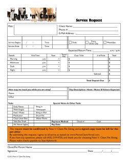


© Copyright 2025