
Document 400047
Van De Graaff: Human
Anatomy, Sixth Edition
IV. Support and Movement
6. Skeletal System:
Introduction and the Axial
Skeleton
Skeletal System:
Introduction
and the Axial Skeleton
© The McGraw−Hill
Companies, 2001
6
Organization of the Skeletal
System 132
Functions of the Skeletal System 134
Bone Structure 135
Bone Tissue 138
Bone Growth 140
Developmental Exposition:
The Axial Skeleton 141
Skull 144
Vertebral Column 158
Rib Cage 164
CLINICAL CONSIDERATIONS 165
Clinical Case Study Answer 168
Important Clinical Terminology 169
Chapter Summary 170
Review Activities 170
Clinical Case Study
A 68-year-old man visited his family doctor for his first physical examination in 30 years. Upon
sensing a disgruntled patient, the doctor gently tried to determine the reason. In response to
the doctor’s inquiry, the patient blurted out, “The nurse who measured my height is incompetent! I know for a fact I used to be six feet even when I was in the Navy, but she tells me I’m
5′10″!” The doctor then performed the measurement himself, noting that although the patient’s posture was excellent, he was indeed 5′10″, just as the nurse had said. He explained to
the patient that the spine contains some nonbony tissue, which shrivels up a bit over the years.
The patient interrupted, stating indignantly that he knew anatomic terms and principles and
would like a detailed explanation. How would you explain the anatomy of the vertebral column
and the changes it undergoes during the aging process?
Hints: The patient’s normal posture and the fact that he had no complaints of pain indicated
good health for his age. Examine figure 6.32 and carefully read the accompanying caption. Also
see the Clinical Considerations section at the end of the chapter.
FIGURE: Decreased height, postural
changes, and loss of bone density are just a
few of the age-related alterations that may
afflict the skeleton.
Van De Graaff: Human
Anatomy, Sixth Edition
Unit 4
132
IV. Support and Movement
6. Skeletal System:
Introduction and the Axial
Skeleton
Support and Movement
ORGANIZATION
OF THE SKELETAL SYSTEM
The axial and appendicular components of the skeletal system of
an adult human consist of 206 individual bones arranged to form a
strong, flexible body framework.
Objective 1
Describe the division of the skeletal system into
axial and appendicular components.
CHAPTER 6
© The McGraw−Hill
Companies, 2001
The adult skeletal system consists of approximately 206 bones.
The exact number of bones differs from person to person depending on age and genetic factors. At birth, the skeleton consists of
about 270 bones. As further bone development (ossification) occurs during infancy, the number increases. During adolescence,
however, the number of bones decreases, as separate bones gradually fuse. Each bone is actually an organ that plays a part in the
total functioning of the skeletal system. The science concerned
with the study of bones is called osteology.
Some adults have extra bones within the sutures (joints) of
the skull called sutural (wormian) bones. Additional bones may
develop in tendons in response to stress as the tendons repeatedly move across a joint. Bones formed this way are called
wormian bone: from Ole Worm, Danish physician, 1588–1654
sesamoid (ses'a˘-moid) bones. Sesamoid bones, like the sutural
bones, vary in number. The patellae (“kneecaps”) are two
sesamoid bones all people have.
For convenience of study, the skeleton is divided into axial
and appendicular portions, as summarized in table 6.1 and shown
in figure 6.1. The axial skeleton consists of the bones that form
the axis of the body and support and protect the organs of the
head, neck, and trunk. The components of the axial skeleton are
as follows:
1. Skull. The skull consists of two sets of bones: the cranial
bones that form the cranium, or braincase, and the facial
bones that support the eyes and nose and form the bony
framework of the oral cavity.
2. Auditory ossicles. Three auditory ossicles (“ear bones”)
are present in the middle-ear chamber of each ear and
serve to transmit sound impulses.
3. Hyoid bone. The hyoid bone is located above the larynx
(“voice box”) and below the mandible (“jawbone”). It supports the tongue and assists in swallowing.
4. Vertebral column. The vertebral column (“backbone”)
consists of 26 individual bones separated by cartilaginous
intervertebral discs. In the pelvic region, several vertebrae
sesamoid: Gk. sesamon, like a sesame seed
TABLE 6.1 Bones of the Adult Skeleton
Axial Skeleton
Appendicular Skeleton
Auditory Ossicles—6 Bones
Pectoral Girdle—5 Bones
14 Facial Bones
8 Cranial Bones
malleus (2)
sternum* (1)
maxilla (2)
frontal (1)
incus (2)
scapula (2)
palatine (2)
parietal (2)
stapes (2)
clavicle (2)
zygomatic (2)
occipital (1)
Hyoid—1 bone
Upper Extremities—60 Bones
lacrimal (2)
temporal (2)
Vertebral Column—26 Bones
humerus (2)
nasal (2)
sphenoid (1)
cervical vertebra (7)
radius (2)
metacarpal bones (10)
vomer (1)
ethmoid (1)
thoracic vertebra (12)
ulna (2)
phalanges (28)
inferior nasal concha (2)
lumbar vertebra (5)
Pelvic Girdle—3 Bones
mandible (1)
sacrum (1) (4 or 5 fused bones)
sacrum* (1)
coccyx (1) (3–5 fused bones)
os coxae (2) (each contains 3 fused bones)
Rib Cage—25 Bones
Lower Extremities—60 Bones
rib (24)
femur (2)
sternum (1)
tibia (2)
metatarsal bones (10)
fibula (2)
phalanges (28)
Skull—22 Bones
carpal bones (16)
tarsal bones (14)
patella (2)
*Although the sternum and sacrum are bones of the axial skeleton, technically speaking they are also considered bones of the pectoral and pelvic girdles, respectively.
Van De Graaff: Human
Anatomy, Sixth Edition
IV. Support and Movement
6. Skeletal System:
Introduction and the Axial
Skeleton
Chapter 6
Frontal bone
Skull
© The McGraw−Hill
Companies, 2001
Skeletal System: Introduction and the Axial Skeleton
133
Parietal bone
Temporal bone
Occipital
bone
Zygomatic bone
Maxilla
Mandible
Clavicle
Sternum
Rib
cage
Pectoral
girdle
Scapula
Costal cartilages
Ribs
Humerus
Vertebral column
Ulna
Ilium
Pubis
Sacrum
CHAPTER 6
Os
coxae
Pelvic
girdle
Coccyx
Ischium
Radius
Carpal bones
Metacarpal bones
Phalanges
Femur
Patella
Tibia
Fibula
Calcaneus
Tarsal bones
Creek
Metatarsal bones
Phalanges
(a)
(b)
FIGURE 6.1 The human skeleton. (a) An anterior view and (b) a posterior view. The axial portion is colored light blue.
Van De Graaff: Human
Anatomy, Sixth Edition
134
Unit 4
IV. Support and Movement
6. Skeletal System:
Introduction and the Axial
Skeleton
Support and Movement
are fused to form the sacrum, which is the attachment portion of the pelvic girdle. A few terminal vertebrae are fused
to form the coccyx (“tailbone”).
5. Rib cage. The rib cage forms the bony and cartilaginous
framework of the thorax. It articulates posteriorly with the
thoracic vertebrae and includes the 12 pairs of ribs, the
flattened sternum, and the costal cartilages that connect the
ribs to the sternum.
CHAPTER 6
© The McGraw−Hill
Companies, 2001
The appendicular (ap''en-dik'yoo-lar) skeleton is composed
of the bones of the upper and lower extremities and the bony girdles that anchor the appendages to the axial skeleton. The components of the appendicular skeleton are as follows:
1. Pectoral girdle. The paired scapulae (“shoulder blades”)
and clavicles (“collarbones”) are the appendicular components of the pectoral girdle, and the sternum (“breastbone”)
is the axial component. The primary function of the pectoral girdle is to provide attachment for the muscles that
move the brachium (arm) and antebrachium (forearm).
2. Upper extremities. Each upper extremity contains a
proximal humerus within the brachium, an ulna and radius
within the antebrachium, the carpal bones, the metacarpal
bones, and the phalanges (“finger bones”) of the hand.
3. Pelvic girdle. The two ossa coxae (“hipbones”) are the
appendicular components of the pelvic girdle, and the
sacrum is the axial component. The ossae coxae are united
anteriorly by the symphysis (sim'f ˘ı -sis) pubis and posteriorly
by the sacrum. The pelvic girdle supports the weight of the
body through the vertebral column and protects the viscera
within the pelvic cavity.
4. Lower extremities. Each lower extremity contains a
proximal femur (“thighbone”) within the thigh, a tibia
(“shinbone”) and fibula within the leg, the tarsal bones, the
metatarsal bones, and the phalanges (“toe bones”) of the
foot. In addition, the patella (pa˘-tel'a˘; “kneecap”) is located
on the anterior surface of the knee joint, between the
thigh and leg.
Knowledge Check
1. List the bones of the body that you can palpate. Indicate
which are bones of the axial skeleton and which are bones
of the appendicular skeleton.
2. What are sesamoid bones and where are they found?
3. Describe the locations and functions of the pectoral and
pelvic girdles.
ossicle: L. ossiculum, little bone
FUNCTIONS
OF THE SKELETAL SYSTEM
The bones of the skeleton perform the mechanical functions of support, protection, and leverage for body movement and the metabolic functions of hemopoiesis and storage of fat and minerals.
Objective 2
Discuss the principal functions of the skeletal
system and identify the body systems served by these functions.
The strength of bone comes from its inorganic components, of
such durability that they resist decomposition even after death.
Much of what we know of prehistoric animals, including humans, has been determined from preserved skeletal remains.
When we think of bone, we frequently think of a hard, dry structure. In fact, the term skeleton comes from a Greek word meaning
“dried up.” Living bone, however, is not inert material; it is dynamic and adaptable. It performs many body functions, including
support, protection, leverage for body movement, hemopoiesis in
the red bone marrow (fig. 6.2), fat storage in the medullary cavity, and mineral storage.
1. Support. The skeleton forms a rigid framework to which
the softer tissues and organs of the body are attached. It is
of interest that the skeleton’s 206 bones support a mass of
muscles and organs that may weigh 5 times as much as the
bones themselves.
2. Protection. The skull and vertebral column enclose the
brain and spinal cord; the rib cage protects the heart,
lungs, great vessels, liver, and spleen; and the pelvic girdle
supports and protects the pelvic viscera. Even the sites
where blood cells are produced are protected within the
spongy bone tissue of certain bones.
3. Body movement. Bones serve as anchoring attachments
for most skeletal muscles. In this capacity, the bones act as
levers (with the joints functioning as pivots) when muscles
contract and cause body movement.
4. Hemopoiesis. The process of blood cell formation is
called hemopoiesis (hem''o˘-poi-e'sis). It takes place in tissue
called red bone marrow located internally in some bones
(fig. 6.2). In an infant, the spleen and liver produce red
blood cells, but as the bones mature, the bone marrow
takes over this formidable task. It is estimated that an average of 2.5 million red blood cells are produced every second by the red bone marrow to replace those that are worn
out and destroyed by the liver.
5. Fat storage. Lipid is stored in the adipose tissue within the
medullary cavity of certain bones. The adipose tissue and its
lipid content are known as yellow bone marrow (fig. 6.2).
6. Mineral storage. The inorganic matrix of bone is composed
primarily of the minerals calcium and phosphorus. These
Van De Graaff: Human
Anatomy, Sixth Edition
IV. Support and Movement
6. Skeletal System:
Introduction and the Axial
Skeleton
© The McGraw−Hill
Companies, 2001
Chapter 6
Skeletal System: Introduction and the Axial Skeleton
135
In summary, the skeletal system is not an isolated body system. It is associated with the muscle system in storing calcium
needed for muscular contraction and providing attachments for
muscles as they span the movable joints. The skeletal system
serves the circulatory system by producing blood cells in protected sites. Directly or indirectly, the skeletal system supports
and protects all of the systems of the body.
Red bone
marrow in
spongy bone
Periosteum
Knowledge Check
4. List the functions of the skeletal system.
5. Discuss two ways in which the skeletal system serves the
circulatory system in the production of blood. What are
two ways in which it serves the muscular system?
Medullary cavity
BONE STRUCTURE
Compact bone
Each bone has a characteristic shape and diagnostic surface features that indicate its functional relationship to other bones, muscles, and to the body structure as a whole.
Objective 3
Classify bones according to their shapes and
give an example of each type.
FIGURE 6.2 Hemopoiesis is the process by which blood cells are
formed. In an adult, blood cells are formed in the red bone marrow.
Objective 4
Describe the various markings on the surfaces
of bones.
Objective 5
minerals which account for approximately two-thirds of the
weight of bone, give bone its firmness and strength. About
95% of the calcium and 90% of the phosphorus within the
body are deposited in the bones and teeth. Although the
concentration of these inorganic salts within the blood is
kept within narrow limits, both are essential for other body
functions. Calcium is necessary for muscle contraction, blood
clotting, and the movement of ions and nutrients across cell
membranes. Phosphorus is required for the activities of the
nucleic acids DNA and RNA, as well as for ATP utilization.
If mineral salts are not present in the diet in sufficient
amounts, they may be withdrawn from the bones until they
are replenished through proper nutrition. In addition to calcium and phosphorus, lesser amounts of magnesium, sodium,
fluorine, and strontium are stored in bone tissue.
Vitamin D assists in the absorption of calcium and phosphorus from the small intestine into the blood. As bones develop
in a child, it is extremely important that the child’s diet contain an
adequate amount of these two minerals and vitamin D. If the diet is
deficient in these essentials, the blood level falls below that necessary for calcification, and a condition known as rickets develops
(see fig. 5.11). Rickets is characterized by soft bones that may result in bowlegs and malformation of the head, chest, and pelvic
girdle.
Describe the gross features of a typical long
bone and list the functions of each surface feature.
The shape and surface features of each bone indicate its functional role in the skeleton (table 6.2). Bones that are long, for
example, provide body support and function as levers during
body movement. Bones that support the body are massive and
have large articular surfaces and processes for muscle attachment.
Roughened areas on these bones may serve for the attachment of
ligaments, tendons, or muscles. A flattened surface provides an
attachment site for a large muscle or may provide protection.
Grooves around an articular end of a bone indicate where a tendon or nerve passes, and openings through a bone permit the
passage of nerves or blood vessels.
Shapes of Bones
The bones of the skeleton are grouped on the basis of shape into
four principal categories: long bones, short bones, flat bones, and irregular bones (fig. 6.3).
1. Long bones. Long bones are longer than they are wide
and function as levers. Most of the bones of the upper and
lower extremities are of this type (e.g., the humerus, radius,
CHAPTER 6
Yellow bone
marrow
Van De Graaff: Human
Anatomy, Sixth Edition
136
Unit 4
IV. Support and Movement
6. Skeletal System:
Introduction and the Axial
Skeleton
© The McGraw−Hill
Companies, 2001
Support and Movement
TABLE 6.2 Surface Features of Bone
Structure
Description and Example
Articulating Surfaces
condyle (kon'dil)
A large, rounded articulating knob (the occipital
condyle of the occipital bone)
facet
A flattened or shallow articulating surface (the
costal facet of a thoracic vertebra)
head
A prominent, rounded articulating end of a bone
(the head of the femur)
Short bone
(i.e., cuboidal bone)
CHAPTER 6
Depressions and Openings
alveolus (al-ve'o˘-lus)
A deep pit or socket (the dental alveoli [tooth
sockets] in the maxilla and mandible)
fissure
A narrow, slitlike opening (the superior orbital
fissure of the sphenoid bone)
foramen (fo˘-ra'men—
plural, foramina)
A rounded opening through a bone (the foramen
magnum of the occipital bone)
fossa (fos'a˘)
A flattened or shallow surface (the mandibular
fossa of the temporal bone)
sinus
A cavity or hollow space in a bone (the frontal
sinus of the frontal bone)
sulcus
A groove that accommodates a vessel, nerve, or
tendon (the intertubercular sulcus of the
humerus)
Nonarticulating Prominences
crest
A narrow, ridgelike projection (the iliac crest of
the os coxae)
epicondyle
A projection adjacent to a condyle (the medial
epicondyle of the femur)
process
Any marked bony prominence (the mastoid
process of the temporal bone)
ramus
A flattened angular part of a bone (the ramus of
the mandible)
spine
A sharp, slender process (the spine of the scapula)
trochanter
A massive process found only on the femur (the
greater trochanter of the femur)
tubercle (too'ber-k'l)
A small, rounded process (the greater tubercle of
the humerus)
tuberosity
A large, roughened process (the radial tuberosity
of the radius)
ulna, metacarpal bones, femur, tibia, fibula, metatarsal
bones, and phalanges).
2. Short bones. Short bones are somewhat cube-shaped and
are found in the wrist and ankle where they transfer forces
of movement.
3. Flat bones. Flat bones have a broad surface for muscle attachment or protection of underlying organs (e.g., the cranial bones, ribs, and bones of the shoulder girdle).
Flat bone
(i.e., parietal bone)
Long bone
(i.e., femur)
Irregular bone
(i.e., thoracic vertebra)
FIGURE 6.3 Examples of bone types, as classified by shape.
4. Irregular bones. Irregular bones have varied shapes and
many surface features for muscle attachment or articulation
(e.g., the vertebrae and certain bones of the skull).
Bone tissue is organized as compact (dense) bone or spongy
(cancellous) bone, and most bones have both types. Compact
bone is hard and dense, and is the protective exterior portion of
all bones. The spongy bone, when it occurs, is deep to the compact bone and is quite porous. The microscopic structure of
spongy and compact bone will be considered shortly.
In a flat bone of the skull, the spongy bone is sandwiched
between the compact bone and is called a diploe (dip'lo-e)
(fig. 6.4). Because of this protective layering of bone tissue, a
blow to the head may fracture the outer compact bone layer
without harming the inner compact bone layer and the brain.
Structure of a Typical Long Bone
The long bones of the skeleton have a descriptive terminology
all their own. In a long bone from an appendage, the bone shaft,
or diaphysis (di-af'ı˘-sis), consists of a cylinder of compact bone
surrounding a central cavity called the medullary (med'yoo-lar-e)
cavity (fig. 6.5). The medullary cavity is lined with a thin layer
facet: Fr. facette, little face
trochanter: Gk. trochanter, runner
tuberosity: L. tuberosus, lump
diploe: Gk. diplous, double
diaphysis: Gk. dia, throughout; physis, growth
Van De Graaff: Human
Anatomy, Sixth Edition
IV. Support and Movement
6. Skeletal System:
Introduction and the Axial
Skeleton
Chapter 6
© The McGraw−Hill
Companies, 2001
Skeletal System: Introduction and the Axial Skeleton
137
CHAPTER 6
FIGURE 6.4 A section through the skull showing diploe. Diploe is
a layer of spongy bone sandwiched between two surface layers of
compact bone. It is extremely strong yet light in weight.
of connective tissue called the endosteum (en-dos'te-um). In an
adult, the cavity contains yellow bone marrow, so named because it contains large amounts of yellow fat. On each end of the
diaphysis is an epiphysis (e˘-pif'e˘-sis), consisting of spongy bone
surrounded by a layer of compact bone. Red bone marrow is
found within the porous chambers of spongy bone. In an adult,
hemopoiesis (the production of blood cells; see chapter 16) occurs in the red bone marrow, especially that of the sternum, vertebrae, portions of the ossa coxae, and the proximal epiphyses of
the femora and humeri. Articular cartilage, which is composed
of thin hyaline cartilage, caps each epiphysis and facilitates joint
movement. Along the diaphysis are nutrient foramina—small
openings into the bone that allow nutrient vessels to pass into
the bone for nourishment of the living tissue.
Between the diaphysis and epiphysis is a cartilaginous epiphyseal (ep''ı˘-fiz'e-al) plate—a region of mitotic activity that is responsible for linear bone growth. As bone growth is completed, an
epiphyseal line replaces the plate and final ossification occurs
between the epiphysis and the diaphysis. A periosteum (per''eos'te-um) of dense regular connective tissue covers the surface of
epiphysis: Gk. epi, upon; physis, growth
periosteum: Gk. peri, around; osteon, bone
FIGURE 6.5 A diagram of a long bone (the humerus) shown in a
partial longitudinal section.
the bone, except over the articular cartilage. This highly vascular
layer serves as a place for a tendon-muscle attachment and is responsible for appositional bone growth (increase in width). The periosteum is secured to the bone by perforating (Sharpey’s) fibers
(fig. 6.5), composed of bundles of collagenous fibers.
Fracture of a long bone in a young person may be especially
serious if it damages an epiphyseal plate. If such an injury goes
untreated, or is not treated properly, longitudinal growth of the bone
may be arrested or slowed, resulting in permanent shortening of the
affected limb.
Sharpey’s fibers: from William Sharpey, Scottish physiologist and histologist,
1802–80
Van De Graaff: Human
Anatomy, Sixth Edition
138
Unit 4
IV. Support and Movement
6. Skeletal System:
Introduction and the Axial
Skeleton
© The McGraw−Hill
Companies, 2001
Support and Movement
Lacuna
Osteoclast
Osteocyte
Osteocyte
Osteoblasts
Canaliculi
(a)
(b)
CHAPTER 6
FIGURE 6.6 (a) Types of bone cells. (b) A photomicrograph of an osteocyte within a lacuna.
Knowledge Check
6. Using examples, discuss the function of each of the four
kinds of bones as determined by shape.
7. Define each of the following surface markings on bones:
condyle, head, facet, process, crest, epicondyle, fossa, alveolus,
foramen, and sinus.
8. Diagram a sagittal view of a typical long bone of a child
and label the diaphysis, medullary cavity, epiphyses, articular cartilages, nutrient foramen, periosteum, and epiphyseal
plates. Explain the function of each of these structures.
secrete unmineralized ground substance. They are abundant in areas
of high metabolism within bone, such as under the periosteum and
bordering the medullary cavity. Osteocytes (os'te-o˘-sı˘ts) are mature
bone cells (figs. 6.6 and 6.7) derived from osteoblasts that have secreted bone tissue around themselves. Osteocytes maintain healthy
bone tissue by secreting enzymes and influencing bone mineral content. Osteoclasts (os'te-o˘-klasts) are large multinuclear cells (fig. 6.6)
that enzymatically break down bone tissue, releasing calcium, magnesium, and other minerals to the blood. These cells are important
in bone growth, remodeling, and healing. Bone-lining cells are derived from osteoblasts along the surface of most bones in the adult
skeleton. These cells are thought to regulate the movement of calcium and phosphate into and out of bone matrix.
BONE TISSUE
Spongy and Compact Bone Tissues
Bone tissue is composed of several types of bone cells embedded
in a matrix of ground substance, inorganic salts (calcium and phosphorus), and collagenous fibers. Bone cells and ground substance
give bone flexibility and strength; the inorganic salts give it hardness.
As mentioned earlier, most bones contain both spongy and compact bone tissues (fig. 6.7). Spongy bone tissue is located deep to
the compact bone tissue, and is quite porous. Minute spikes of
bone tissue, called trabeculae (tra˘-bek'yu˘-le), give spongy bone a
latticelike appearance. Spongy bone is highly vascular and provides great strength to bone with minimal weight.
Compact bone tissue forms the external portion of a
bone and is very hard and dense. It consists of precise arrangements of microscopic cylindrical structures oriented parallel to
the long axis of the bone (fig. 6.7). These columnlike structures are the osteons (os'te-onz), or haversian systems, of the
bone tissue. The matrix of an osteon is laid down in concentric rings, called lamellae (la˘-mel'e), that surround a central
(haversian) canal (fig. 6.8). The central canal contains minute
Objective 6
Identify the five types of bone cells and list the
functions of each.
Objective 7
Distinguish between spongy and compact
bone tissues.
Bone Cells
There are five principal types of bone cells contained within bone
tissue. Osteogenic (os''te-o˘-jen'ik) cells are found in the bone tissues
in contact with the endosteum and the periosteum. These cells respond to trauma, such as a fracture, by giving rise to bone-forming
cells (osteoblasts) and bone-destroying cells (osteoclasts). Osteoblasts
(os'te-o˘-blasts) are bone-forming cells (fig. 6.6) that synthesize and
osteoblast: Gk. osteon, bone; blastos, offspring or germ
osteoclast: GK. osteon, bone; klastos, broken
haversian system: from Clopton Havers, English anatomist, 1650–1702
Van De Graaff: Human
Anatomy, Sixth Edition
IV. Support and Movement
6. Skeletal System:
Introduction and the Axial
Skeleton
Chapter 6
Periosteum
© The McGraw−Hill
Companies, 2001
Skeletal System: Introduction and the Axial Skeleton
External circumferential lamellae
Arteriole
Central canal
139
Venule
Nerve
Central
canal
Canaliculi
Lamellae
Medullary
cavity
(c)
(a)
Osteocyte
Lacuna
CHAPTER 6
Perforating
fibers
Canaliculi
Blood vessels
Perforating
canals
Central
canal
Trabeculae of
spongy bone
(d)
(b)
FIGURE 6.7 Compact bone tissue. (a) A diagram of the femur showing a cut through the compact bone into the medullary cavity. (b) The
arrangement of the osteons within the diaphysis of the bone. (c) An enlarged view of an osteon showing the osteocytes within lacunae and the
concentric lamellae. (d) An osteocyte within a lacuna.
LA
CA
Canaliculi
Osteocyte
within a
lacuna
Central
canal
Lamella
CA
LA
(a)
(b)
FIGURE 6.8 Bone tissue as seen in (a) a scanning electron micrograph and (b) a photomicrograph. The lacunae (LA) provides spaces for the
osteocytes, which are connected to one another by canaliculi (CA). Note the divisions between the lamellae (see arrows).
Van De Graaff: Human
Anatomy, Sixth Edition
140
Unit 4
IV. Support and Movement
6. Skeletal System:
Introduction and the Axial
Skeleton
© The McGraw−Hill
Companies, 2001
Support and Movement
(a)
(b)
CHAPTER 6
(c)
(d)
(e)
(f)
FIGURE 6.9 The growth process of a long bone, beginning with (a) the cartilaginous model as it occurs in an embryo at 6 weeks. The bone
develops (b–e) through intermediate stages to (f ) adult bone.
nutrient vessels and a nerve. Osteocytes within spaces called
lacunae (la˘-kyoo'ne) are regularly arranged between the lamellae. The lacunae are connected by tiny channels called canaliculi (kan'' a˘-lik'yu˘-li), through which nutrients diffuse.
Metabolic activity within bone tissue occurs at the osteon
level. Between osteons there are incomplete remnants of osteons, called interstitial systems. Perforating (Volkmann’s)
canals penetrate compact bone, connecting osteons with blood
vessels and nerves.
Knowledge Check
9. Construct a sample table listing the location and function
of each type of cell found within bone tissue
10. Define osteon and sketch the arrangement of osteons
within compact bone tissue.
Volkmann’s canal: from Alfred Volkmann, German physiologist, 1800–77
BONE GROWTH
The development of bone from embryo to adult depends on the orderly processes of cell division, growth, and ongoing remodeling.
Bone growth is influenced by genetics, hormones, and nutrition.
Objective 8
Describe the process of endochondral
ossification as related to bone growth.
In most bone development, a cartilaginous model is gradually replaced by bone tissue during endochondral bone formation (see Developmental Exposition, pp. 141–142). As the cartilage model
grows, the chondrocytes (cartilage cells) in the center of the shaft
hypertrophy, and minerals are deposited within the matrix in a
process called calcification (fig. 6.9). Calcification restricts the passage of nutrients to the chondrocytes, causing them to die. At the
same time, some cells of the perichondrium (dense regular connective tissue surrounding cartilage) differentiate into osteoblasts.
These cells secrete osteoid (os'te-oid), the hardened organic
Van De Graaff: Human
Anatomy, Sixth Edition
IV. Support and Movement
6. Skeletal System:
Introduction and the Axial
Skeleton
© The McGraw−Hill
Companies, 2001
Developmental Exposition
The Axial Skeleton
certain bones of the cranium are formed this way. Sesamoid bones
are specialized intramembranous bones that develop in tendons.
The patella is an example of a sesamoid bone.
EXPLANATION
DEVELOPMENT OF THE SKULL
Development of Bone
Bone formation, or ossification, begins at about the fourth week of
embryonic development, but ossification centers cannot be readily observed until about the tenth week (exhibit I). Bone tissue
derives from specialized migratory cells of mesoderm (see
fig. 4.13) known as mesenchyme. Some of the embryonic mesenchymal cells will transform into chondroblasts (kon'dro-blasts)
and develop a cartilage matrix that is later replaced by bone in a
process known as endochondral (en''do˘-kon'dral) ossification.
Most of the skeleton is formed in this fashion—first it goes
through a hyaline cartilage stage and then it is ossified as bone.
A smaller number of mesenchymal cells develop into bone
directly, without first going through a cartilage stage. This type
of bone-formation process is referred to as intramembranous
(in''tra˘-mem'bra˘-nus) ossification. The clavicles, facial bones, and
chondroblast: Gk. chondros, cartilage; blastos, offspring or germ
The formation of the skull is a complex process that begins during the fourth week of embryonic development and continues
well beyond the birth of the baby. Three aspects of the embryonic skull are involved in this process: the chondrocranium, the
neurocranium, and the viscerocranium (exhibit II). The chondrocranium is the portion of the skull that undergoes endochondral ossification to form the bones supporting the brain. The
neurocranium is the portion of the skull that develops through
membranous ossification to form the bones covering the brain
and facial region. The viscerocranium (splanchnocranium) is the
portion that develops from the embryonic visceral arches to form
the mandible, auditory ossicles, the hyoid bone, and specific
processes of the skull.
chondrocranium: Gk. chondros, cartilage; kranion, skull
viscerocranium: L. viscera, soft parts; Gk. kranion, skull
Parietal
bones
Occipital
bone
Frontal
bones
Temporal
bone
Humerus
Zygomatic
bone
Maxilla
Nasal bone
Mandible
Metacarpal
bones
Phalanges
Carpal bones
Ribs
Radius
Ulna
Chondrocranium
Vertebrae
Clavicle
Scapula
Femur
Tibia
Fibula
Ilium
Sacrum
Coccyx
(a)
Phalanges
Metatarsal bones
Tarsal bones
Creek
(b)
EXHIBIT I Ossification centers of the skeleton of a 10-week-old fetus. (a) The diagram depicts endochondrial ossification in red and intramembranous ossification in a stippled pattern. The cartilaginous portions of the skeleton are shown in gray. (b) The photograph shows
the ossification centers stained with a red indicator dye.
141
Van De Graaff: Human
Anatomy, Sixth Edition
IV. Support and Movement
6. Skeletal System:
Introduction and the Axial
Skeleton
© The McGraw−Hill
Companies, 2001
(continued)
EXHIBIT II The embryonic skull at 12 weeks is composed of bony elements from three developmental sources: the chondrocranium
(colored blue-gray), the neurocranium (colored light yellow), and the viscerocranium (colored salmon).
component of bone. As the perichondrium calcifies, it gives rise to
a thin plate of compact bone called the periosteal bone collar.
The periosteal bone collar is surrounded by the periosteum.
A periosteal bud, consisting of osteoblasts and blood vessels, invades the disintegrating center of the cartilage model from
the periosteum. Once in the center, the osteoblasts secrete osteoid, and a primary ossification center is established. Ossification then expands into the deteriorating cartilage. This process is
repeated in both the proximal and distal epiphyses, forming secondary ossification centers where spongy bone develops.
Once the secondary ossification centers have been formed,
bone tissue totally replaces cartilage tissue, except at the articular
ends of the bone and at the epiphyseal plates. An epiphyseal
plate contains five histological zones (fig. 6.10). The reserve zone
(zone of resting cartilage) borders the epiphysis and consists of
small chondrocytes irregularly dispersed throughout the intercellular matrix. The chondrocytes in this zone anchor the epiphyseal plate to the bony epiphysis. The proliferation zone (zone of
proliferating cartilage) consists of larger, regularly arranged chondrocytes that are constantly dividing. The hypertrophic zone (zone
142
Epiphyseal border
Reserve
zone
Proliferation
zone
Chondrocytes
Hypertrophic
zone
Epiphyseal border
Resorption
zone
Bone tissue
Ossification
zone
Red bone
marrow
Diaphyseal border
FIGURE 6.10 A photomicrograph from an epiphyseal plate (63×).
Van De Graaff: Human
Anatomy, Sixth Edition
IV. Support and Movement
6. Skeletal System:
Introduction and the Axial
Skeleton
Chapter 6
© The McGraw−Hill
Companies, 2001
Skeletal System: Introduction and the Axial Skeleton
143
TABLE 6.3 Average Age of Completion
of Bone Ossification
Bone
Chronological
Age of Fusion
Scapula
18–20
Clavicle
23–31
Bones of upper extremity
17–20
Os coxae
18–23
Bones of lower extremity
18–22
Vertebra
25
Sacrum
23–25
Sternum (body)
23
Sternum (manubrium, xiphoid)
30+
Diaphysis
Epiphyseal
plate
Epiphysis
Epiphyseal
plates
diograph of a child’s hand. The plates indicate that the bones are still
growing in length.
The time at which epiphyseal plates ossify varies greatly from
bone to bone, but it usually occurs between the ages of 18 and
20 within the long bones (table 6.3). Because ossification of the
epiphyseal cartilages within each bone occurs at predictable times,
radiologists can determine the ages of people who are still growing
by examining radiographs of their bones (fig. 6.11). Large discrepancies between bone age and chronological age may indicate a genetic or endocrine abnormality.
Bone is continually being remodeled over the course of a
person’s life. Bony prominences develop as stress is applied to the
periosteum, causing the osteoblasts to secrete osteoid and form
new bone tissue. The greater trochanter of the femur, for example, develops in response to forces of stress applied to the periosteum where the tendons of muscles attach (fig. 6.12). Even
though a person has stopped growing in height, bony processes
may continue to enlarge somewhat if he or she remains physically active.
Spongy bone
Compact bone
Medullary cavity
Creek
FIGURE 6.12 A longitudinal section of the proximal end of a
femur showing stress lines within spongy bone.
CHAPTER 6
FIGURE 6.11 The presence of epiphyseal plates as seen in a raof hypertrophic cartilage) consists of very large chondrocytes
that are arranged in columns. The linear growth of long bones is
due to the cellular proliferation at the proliferation zone and the
growth and maturation of these new cells within the hypertrophic zone. The resorption zone (zone of dechondrification) is
the area where a change in mineral content is occurring. The ossification zone (zone of calcified cartilage) is a region of transformation from cartilage tissue to bone tissue. The chondrocytes
within this zone die because the intercellular matrix surrounding
them becomes calcified. Osteoclasts then break down the calcified matrix and the area is invaded by osteoblasts and capillaries
from the bone tissue of the diaphysis. As the osteoblasts mature,
osteoid is secreted and bone tissue is formed. The result of this
process is a gradual increase in the length of the bone at the
epiphyseal plates.
Van De Graaff: Human
Anatomy, Sixth Edition
144
Unit 4
IV. Support and Movement
6. Skeletal System:
Introduction and the Axial
Skeleton
Support and Movement
As new bone layers are deposited on the outside surface of
the bone, osteoclasts dissolve bone tissue adjacent to the
medullary cavity. In this way, the size of the cavity keeps pace
with the increased growth of the bone.
Even the absence of stress causes a remodeling of bones.
This effect can best be seen in the bones of bedridden or paralyzed individuals. Radiographs of their bones reveal a marked loss
of bone tissue. The absence of gravity that accompanies space
flight may result in mineral loss from bones if an exercise regimen is not followed.
The movement of teeth in orthodontics involves bone remodeling. The dental alveoli (tooth sockets) are reshaped through the
activity of osteoclast and osteoblast cells as stress is applied with
braces. The use of traction in treating certain skeletal disorders has a
similar effect.
CHAPTER 6
Knowledge Check
11. List the zones of an epiphyseal plate and briefly describe
the characteristics of each.
12. Explain the function of osteoblasts and osteoclasts in endochondral ossification and bone growth.
SKULL
The human skull, consisting of 8 cranial and 14 facial bones, contains several cavities that house the brain and sensory organs.
Each bone of the skull articulates with the adjacent bones and
has diagnostic and functional processes, surface features, and
foramina.
Objective 9
© The McGraw−Hill
Companies, 2001
List the fontanels and discuss their functions.
ducts into the nasal cavity. Middle- and inner-ear cavities are
positioned inferior to the cranial cavity and house the organs of
hearing and balance. The two orbits for the eyeballs are formed
by facial and cranial bones. The oral, or buccal (buk'al) cavity
(mouth), which is only partially formed by bone, is completely
within the facial region (see fig. 2.23).
During fetal development and infancy, the bones of the
cranium are separated by fibrous unions. There are also six large
areas of connective tissue membrane that cover the gaps between
the developing bones. These membranous sheets are called
fontanels (fon''ta˘-nelz'), meaning “little fountains.” The name
derives from the fact that a baby’s pulse can be felt surging in
these “soft spots” on the skull. The fontanels permit the skull to
undergo changes in shape, called molding, during parturition
(childbirth), and they accommodate the rapid growth of the
brain during infancy. Ossification of the fontanels is normally
complete by 20 to 24 months of age. The fontanels are illustrated
in figure 6.13 and briefly described below.
1. Anterior (frontal) fontanel. The anterior fontanel is
diamond-shaped and is the most prominent. It is located
on the anteromedian portion of the skull.
2. Posterior (occipital) fontanel. The posterior fontanel is
positioned at the back of the skull on the median line. It is
also diamond-shaped, but smaller than the anterior
fontanel.
3. Anterolateral (sphenoid) fontanels. The paired anterolateral fontanels are found on both sides of the skull, directly lateral to the anterior fontanel. They are relatively
small and irregularly shaped.
4. Posterolateral (mastoid) fontanels. The paired posterolateral fontanels, also irregularly shaped, are located on the
posterolateral sides of the skull.
Objective 10
Identify the cranial and facial bones of the
skull and describe their structural characteristics.
Objective 11
Describe the location of each of the bones
of the skull and identify the articulations that affix one to
the other.
The skull consists of cranial bones and facial bones. The eight
bones of the cranium articulate firmly with one another to enclose and protect the brain and sensory organs. The 14 facial
bones form the framework for the facial region and support the
teeth. Variation in size, shape, and density of the facial bones is
a major contributor to the individuality of each human face.
The facial bones, with the exception of the mandible (“jawbone”), are also firmly interlocked with one another and the
cranial bones.
The skull has several cavities. The cranial cavity is the
largest, with an approximate capacity of 1,300 to 1,350 cc. The
nasal cavity is formed by both cranial and facial bones and is
partitioned into two chambers, or nasal fossae, by a nasal septum of bone and cartilage. Four sets of paranasal sinuses, located
within the bones surrounding the nasal area, communicate via
During normal childbirth, the fetal skull comes under tremendous pressure. Bones may even shift, altering the shape of the
skull. A common occurrence during molding of the fetal skull is
for the occipital bone to be repositioned under the two parietal
bones. In addition, one parietal bone may shift so as to overlap the other. This makes delivery easier for the mother. If a baby is
born breech (buttocks first), these shifts do not occur. Delivery becomes much more difficult, often requiring the use of forceps.
A prominent sagittal suture extends the anteroposterior
median length of the skull between the anterior and posterior
fontanels. A coronal suture extends from the anterior fontanel
to the anterolateral fontanel. A lambdoid suture extends from
the posterior fontanel to the posterolateral fontanel. A squamous suture connects the posterolateral fontanel to the anterolateral fontanel.
The bones of the skull contain numerous foramina (see
table 6.2) to accommodate nerves, vessels, and other structures.
The foramina of the skull are summarized in table 6.4. Various
fontanel: Fr. fontaine, little fountain
lambdoid: Gk. lambda, letter λ in Greek alphabet
Van De Graaff: Human
Anatomy, Sixth Edition
IV. Support and Movement
6. Skeletal System:
Introduction and the Axial
Skeleton
Chapter 6
© The McGraw−Hill
Companies, 2001
Skeletal System: Introduction and the Axial Skeleton
145
Anterior fontanel
Coronal suture
Parietal bone
Frontal bone
Posterior fontanel
Squamous suture
Anterolateral
fontanel
Lambdoid suture
Nasal bone
Occipital bone
Sphenoid bone
Posterolateral
fontanel
Zygomatic bone
Maxilla
Temporal bone
Mandible
(a)
Frontal bone
Sagittal suture
Parietal bone
ee
Cr
Posterior fontanel
k
Occipital bone
(b)
FIGURE 6.13 The fetal skull showing the six fontanels and the sutures. (a) A right lateral view and (b) a superior view.
views of the skull are shown in figures 6.14 through 6.21; radiographs are shown in figure 6.22.
Although the hyoid bone and the three paired auditory ossicles are not considered part of the skull, they are associated
with it. These bones are described in this section, immediately
following the discussion of the facial bones.
Cranial Bones
The cranial bones enclose and protect the brain and associated
sensory organs. They consist of one frontal, two parietals, two
temporals, one occipital, one sphenoid, and one ethmoid.
Frontal Bone
The frontal bone forms the anterior roof of the cranium, the forehead,
the roof of the nasal cavity, and the superior arches of the orbits,
which contain the eyeballs. The bones of the orbit are summarized in
table 6.5. The frontal bone develops in two halves that grow together.
Generally, they are completely fused by age 5 or 6. A suture sometimes persists between these two portions beyond age 6 and is referred
to as a metopic (me˘-top'ik) suture. The supraorbital margin is a
cranium: Gk. kranion, skull
metopic suture: Gk. metopon, forehead; L. sutura, sew
CHAPTER 6
Anterior fontanel
Coronal suture
Van De Graaff: Human
Anatomy, Sixth Edition
146
Unit 4
IV. Support and Movement
6. Skeletal System:
Introduction and the Axial
Skeleton
© The McGraw−Hill
Companies, 2001
Support and Movement
CHAPTER 6
TABLE 6.4 Major Foramina of the Skull
Foramen
Location
Structures Transmitted
Carotid canal
Petrous part of temporal bone
Internal carotid artery and sympathetic nerves
Greater palatine foramen
Palatine bone of hard palate
Greater palatine nerve and descending palatine vessels
Hypoglossal canal
Anterolateral edge of occipital condyle
Hypoglossal nerve and branch of ascending pharyngeal
artery
Incisive foramen
Anterior region of hard palate, posterior to incisors
Branches of descending palatine vessels and nasopalatine
nerve
Inferior orbital fissure
Between maxilla and greater wing of sphenoid bone
Maxillary nerve of trigeminal cranial nerve, zygomatic
nerve, and infraorbital vessels
Infraorbital foramen
Inferior to orbit in maxilla
Infraorbital nerve and artery
Jugular foramen
Between petrous part of temporal and occipital bones,
posterior to carotid canal
Internal jugular vein; vagus, glossopharyngeal, and accessory
nerves
Foramen lacerum
Between petrous part of temporal and sphenoid bones
Branch of ascending pharyngeal artery and internal carotid
artery
Lesser palatine foramen
Posterior to greater palatine foramen in hard palate
Lesser palatine nerves
Foramen magnum
Occipital bone
Union of medulla oblongata and spinal cord, meningeal
membranes, and accessory nerves; vertebral and spinal
arteries
Mandibular foramen
Medial surface of ramus of mandible
Inferior alveolar nerve and vessels
Mental foramen
Below second premolar on lateral side of mandible
Mental nerve and vessels
Nasolacrimal canal
Lacrimal bone
Nasolacrimal (tear) duct
Cribriform foramina
Cribriform plate of ethmoid bone
Olfactory nerves
Optic foramen
Back of orbit in lesser wing of sphenoid bone
Optic nerve and ophthalmic artery
Foramen ovale
Greater wing of sphenoid bone
Mandibular nerve (branch) of trigeminal nerve
Foramen rotundum
Within body of sphenoid bone
Maxillary nerve (branch) of trigeminal nerve
Foramen spinosum
Posterior angle of sphenoid bone
Middle meningeal vessels
Stylomastoid foramen
Between styloid and mastoid processes of temporal bone
Facial nerve and stylomastoid artery
Superior orbital fissure
Between greater and lesser wings of sphenoid bone
Four cranial nerves (oculomotor, trochlear, ophthalmic
nerve of trigeminal, and abducens)
Supraorbital foramen
Supraorbital ridge of orbit
Supraorbital nerve and artery
Zygomaticofacial foramen
Anterolateral surface of zygomatic bone
Zygomaticofacial nerve and vessels
prominent bony ridge over the orbit. Slightly medial to its midpoint
is an opening called the supraorbital foramen, which provides passage for a nerve, artery, and veins.
The frontal bone also contains frontal sinuses, which are
connected to the nasal cavity (fig 6.22). These sinuses, along
with the other paranasal sinuses, lessen the weight of the skull
and act as resonance chambers for voice production.
Parietal Bone
The two parietal bones form the upper sides and roof of the cranium (figs. 6.15 and 6.17). The coronal suture separates the
frontal bone from the parietal bones, and the sagittal suture
along the superior midline separates the right and left parietals
from each other. The inner concave surface of each parietal
bone, as well as the inner concave surfaces of other cranial
bones, is marked by shallow impressions from convolutions of the
brain and vessels serving the brain.
Temporal Bone
The two temporal bones form the lower sides of the cranium
(figs. 6.15, 6.16, 6.17, and 6.23). Each temporal bone is joined to
its adjacent parietal bone by the squamous suture. Structurally,
each temporal bone has four parts.
1. Squamous part. The squamous part is the flattened plate
of bone at the sides of the skull. Projecting forward is a zygomatic (zi''go-mat'ik) process that forms the posterior portion of the zygomatic arch. On the inferior surface of the
squamous part is the cuplike mandibular fossa, which
forms a joint with the condyle of the mandible. This articulation is the temporomandibular joint.
zygomatic: Gk. zygoma, yolk
Van De Graaff: Human
Anatomy, Sixth Edition
IV. Support and Movement
6. Skeletal System:
Introduction and the Axial
Skeleton
Chapter 6
© The McGraw−Hill
Companies, 2001
Skeletal System: Introduction and the Axial Skeleton
147
Frontal bone
Parietal bone
Temporal bone
Lacrimal bone
Nasal bone
Zygomatic
bone
Inferior nasal concha
Maxilla
CHAPTER 6
Vomer
Mandible
FIGURE 6.14 An anterior view of the skull.
Coronal suture
Parietal bone
Frontal bone
Lambdoid suture
Sphenoid bone
Squamous suture
Ethmoid bone
Temporal bone
Lacrimal bone
Occipital bone
Nasal bone
Zygomatic bone
External acoustic meatus
Infraorbital foramen
Mastoid process
Maxilla
Coronoid process
of mandible
Condylar process
of mandible
Styloid process
Zygomatic process
Mental foramen
Mandibular notch
Angle of mandible
FIGURE 6.15 A lateral view of the skull.
Mandible
Creek
Van De Graaff: Human
Anatomy, Sixth Edition
148
Unit 4
IV. Support and Movement
6. Skeletal System:
Introduction and the Axial
Skeleton
© The McGraw−Hill
Companies, 2001
Support and Movement
Incisors
Canine
Premolars
Incisive foramen
Molars
Median palatine suture
Zygomatic bone
Palatine process of maxilla
Palatine bone
Sphenoid bone
Greater palatine foramen
Zygomatic process
Medial and lateral
pterygoid processes
of sphenoid bone
Vomer
Foramen ovale
Mandibular fossa
Foramen lacerum
Carotid canal
External acoustic meatus
Jugular fossa
CHAPTER 6
Styloid process
Stylomastoid foramen
Mastoid process
Foramen magnum
Occipital condyle
Mastoid foramen
Temporal bone
Parietal bone
Superior nuchal line
Condyloid canal
Occipital bone
External occipital protuberance
Creek
FIGURE 6.16 An inferior view of the skull.
Parietal bone
Frontal
bone
Temporal bone
Occipital bone
Nasal bone
Maxilla
Mandible
Vomer
FIGURE 6.17 A sagittal view of the skull.
Palatine bone
Van De Graaff: Human
Anatomy, Sixth Edition
IV. Support and Movement
6. Skeletal System:
Introduction and the Axial
Skeleton
Chapter 6
© The McGraw−Hill
Companies, 2001
Skeletal System: Introduction and the Axial Skeleton
149
Squamous suture
Supraorbital margin
Mandibular condyle
Mandibular fossa
Zygomatic arch
External acoustic meatus
Coronoid process of
mandible
Mastoid process
of temporal bone
Styloid process
of temporal bone
Ramus of mandible
Jugular foramen
Mental protuberance
Lambdoid suture
Angle of mandible
Occipitomastoid suture
Digastric fossa
Condyloid canal
Occipital condyle
Mandibular foramen
Foramen magnum
CHAPTER 6
FIGURE 6.18 An inferolateral view of the skull.
Frontal bone
Sphenoid bone
Temporal bone
Parietal
bone
Occipital bone
FIGURE 6.19 The floor of the cranial cavity.
Van De Graaff: Human
Anatomy, Sixth Edition
150
Unit 4
IV. Support and Movement
6. Skeletal System:
Introduction and the Axial
Skeleton
© The McGraw−Hill
Companies, 2001
Support and Movement
Frontal
bone
Ethmoid bone
Zygomatic bone
Middle nasal concha
CHAPTER 6
Maxilla
Inferior nasal concha
Vomer
FIGURE 6.20 A posterior view of a frontal (coronal) section of the skull.
Frontal bone
Nasal bone
Lacrimal bone
Zygomatic
bone
Maxilla
FIGURE 6.21 Bones of the orbit.
Van De Graaff: Human
Anatomy, Sixth Edition
IV. Support and Movement
6. Skeletal System:
Introduction and the Axial
Skeleton
Chapter 6
© The McGraw−Hill
Companies, 2001
Skeletal System: Introduction and the Axial Skeleton
Ethmoidal
sinus
151
Frontal sinus
Sphenoidal sinus
Frontal
sinus
Ethmoidal sinuses
Sphenoidal
sinus
Maxillary sinus
Maxillary
sinus
(a)
(b)
TABLE 6.5 Bones Forming the Orbit
Region of the Orbit
Contributing Bones
Roof (superior)
Frontal bone; lesser wing of sphenoid bone
Floor (inferior)
Maxilla; zygomatic bone; palatine bone
Lateral wall
Zygomatic bone
Posterior wall
Greater wing of sphenoid bone
Medial wall
Maxilla; lacrimal bone; ethmoid bone
Superior margin
Frontal bone
Lateral margin
Zygomatic bone
Medial margin
Maxilla
2. Tympanic part. The tympanic part of the temporal bone
contains the external acoustic meatus (me-a'tus), or ear
canal, which is posterior to the mandibular fossa. A thin,
pointed styloid process (figs. 6.16, 6.17, and 6.18) projects
inferiorly from the tympanic part.
3. Mastoid part. The mastoid process, a rounded projection
posterior to the external acoustic meatus, accounts for the
mass of the mastoid part. The mastoid foramen (fig. 6.16)
is directly posterior to the mastoid process. The stylomastoid foramen, located between the mastoid and styloid
processes (fig. 6.16), provides the passage for part of the facial nerve.
styloid: Gk. stylos, pillar
mastoid: Gk. mastos, breast
4. Petrous part. The petrous (pet'rus) part can be seen in the
floor of the cranium (figs. 6.19 and 6.23). The structures of
the middle ear and inner ear are housed in this dense part of
the temporal bone. The carotid (ka˘-rot'id) canal and the
jugular foramen border on the medial side of the petrous
part at the junction of the temporal and occipital bones.
The carotid canal allows blood into the brain via the internal carotid artery, and the jugular foramen lets blood drain
from the brain via the internal jugular vein. Three cranial
nerves also pass through the jugular foramen (see table 6.4).
The mastoid process of the temporal bone can be easily palpated as a bony knob immediately behind the earlobe. This
process contains a number of small air-filled spaces called mastoid
cells that can become infected in mastoiditis, as a result, for example, of a prolonged middle-ear infection.
Occipital Bone
The occipital bone forms the posterior and most of the base of
the skull. It articulates with the parietal bones at the lambdoid
suture. The foramen magnum is the large hole in the occipital
bone through which the spinal cord passes to attach to the brain
stem. On each side of the foramen magnum are the occipital
condyles (fig. 6.16), which articulate with the first vertebra (the
atlas) of the vertebral column. At the anterolateral edge of the
occipital condyle is the hypoglossal canal (fig. 6.17), through
which the hypoglossal nerve passes. A condyloid (kon'd˘-loid)
ı
canal lies posterior to the occipital condyle (fig. 6.16). The
petrous: Gk. petra, rock
magnum: L. magnum, great
CHAPTER 6
FIGURE 6.22 Radiographs of the skull showing the paranasal sinuses. (a) An anteroposterior view and (b) a right lateral view.
Van De Graaff: Human
Anatomy, Sixth Edition
CHAPTER 6
152
Unit 4
IV. Support and Movement
6. Skeletal System:
Introduction and the Axial
Skeleton
© The McGraw−Hill
Companies, 2001
Support and Movement
(a)
(b)
FIGURE 6.23 The temporal bone. (a) A lateral view and (b) a medial view.
external occipital protuberance is a prominent posterior projection on the occipital bone that can be felt as a definite bump just
under the skin. The superior nuchal (noo'kal) line is a ridge of
bone extending laterally from the occipital protuberance to the
mastoid part of the temporal bone. Sutural bones are small clusters of irregularly shaped bones that frequently occur along the
lambdoid suture.
Sphenoid Bone
The sphenoid (sfe'noid) bone forms part of the anterior base
of the cranium and can be viewed laterally and inferiorly
(figs. 6.15 and 6.16). This bone has a somewhat mothlike shape
(fig. 6.24). It consists of a body and laterally projecting greater
and lesser wings that form part of the orbit. The wedgelike
body contains the sphenoidal sinuses and a prominent saddlelike depression, the sella turcica (sel'a˘ tur'sı˘-ka˘). Commonly
called “Turk’s saddle,” the sella turcica houses the pituitary
gland. A pair of pterygoid (ter'ı˘-goid) processes project inferiorly from the sphenoid bone and help form the lateral walls of
the nasal cavity.
Several foramina (figs. 6.16, 6.19, and 6.24) are associated
with the sphenoid bone.
nuchal: Fr. nuque, nape of neck
sphenoid: Gk. sphenoeides, wedgelike
1. The optic canal is a large opening through the lesser wing
into the back of the orbit that provides passage for the
optic nerve and the ophthalmic artery.
2. The superior orbital fissure is a triangular opening between the wings of the sphenoid bone that provides passage for the ophthalmic nerve, a branch of the trigeminal
nerve, and for the oculomotor, trochlear, and abducens
nerves.
3. The foramen ovale is an opening at the base of the lateral
pterygoid plate, through which the mandibular nerve passes.
4. The foramen spinosum is a small opening at the posterior
angle of the sphenoid bone that provides passage for the
middle meningeal vessels.
5. The foramen lacerum (las'er-um) is an opening between
the sphenoid and the petrous part of the temporal bone,
through which the internal carotid artery and the
meningeal branch of the ascending pharyngeal artery pass.
6. The foramen rotundum is an opening just posterior to the
superior orbital fissure, at the junction of the anterior and
medial portions of the sphenoid bone. The maxillary nerve
passes through this foramen.
Located on the inferior side of the cranium, the sphenoid bone
would seem to be well protected from trauma. Actually, just the
opposite is true—and in fact the sphenoid is the most frequently fractured bone of the cranium. It has several broad, thin, platelike extensions that are perforated by numerous foramina. A blow to almost any
portion of the skull causes the buoyed, fluid-filled brain to rebound
against the vulnerable sphenoid bone, often causing it to fracture.
Van De Graaff: Human
Anatomy, Sixth Edition
IV. Support and Movement
6. Skeletal System:
Introduction and the Axial
Skeleton
Chapter 6
© The McGraw−Hill
Companies, 2001
Skeletal System: Introduction and the Axial Skeleton
153
CHAPTER 6
FIGURE 6.24 The sphenoid bone. (a) A superior view and (b) a posterior view.
Ethmoid Bone
The ethmoid bone is located in the anterior portion of the floor
of the cranium between the orbits, where it forms the roof of
the nasal cavity (figs. 6.17, 6.20, and 6.25). An inferior projection of the ethmoid bone, called the perpendicular plate, forms
the superior part of the nasal septum that separates the nasal
cavity into two chambers. Each chamber of the nasal cavity is
referred to as a nasal fossa. Flanking the perpendicular plate on
each side is a large but delicate mass of bone riddled with ethmoidal air cells, collectively constituting the ethmoid sinus. A
spine of the perpendicular plate, the crista galli (kris'ta˘ gal'e),
projects superiorly into the cranial cavity and serves as an attachment for the meninges covering the brain. On both lateral
walls of the nasal cavity are two scroll-shaped plates of the ethmoid bone, the superior and middle nasal conchae (kong'ke—
singular, concha) (fig. 6.26), also known as turbinates. At right
angles to the perpendicular plate, within the floor of the cranium, is the cribriform (krib'rı˘-form) plate, which has numerous cribriform foramina for the passage of olfactory nerves
from the epithelial lining of the nasal cavity. The bones of the
nasal cavity are summarized in table 6.6.
Creek
Cribriform plate
Cribriform foramina
Orbital plate
Ethmoidal air cells
ethmoid: Gk. ethmos, sieve
crista galli: L. crista, crest; galli, cock’s comb
conchae: L. conchae, shells
cribriform: L. cribrum, sieve; forma, like
Crista galli
Superior nasal concha
Middle nasal concha
Perpendicular plate
FIGURE 6.25 An anterior view of the ethmoid bone.
Van De Graaff: Human
Anatomy, Sixth Edition
CHAPTER 6
154
Unit 4
IV. Support and Movement
6. Skeletal System:
Introduction and the Axial
Skeleton
© The McGraw−Hill
Companies, 2001
Support and Movement
FIGURE 6.26 The lateral wall of the nasal cavity.
TABLE 6.6 Bones That Enclose
the Nasal Cavity
Region of
Nasal Cavity
Contributing Bones
Roof (superior)
Ethmoid bone (cribriform plate); frontal bone
Floor (inferior)
Maxilla; palatine bone
Lateral wall
Maxilla; palatine bone
Nasal septum (medial)
Ethmoid bone (perpendicular plate); vomer;
nasal bone
Bridge
Nasal bone
Conchae
Ethmoid bone (superior and middle conchae);
inferior nasal concha
The moist, warm vascular lining within the nasal cavity is susceptible to infections, particularly if a person is not in good
health. Infections of the nasal cavity can spread to several surrounding areas. The paranasal sinuses connect to the nasal cavity and
are especially prone to infection. The eyes may become reddened
and swollen during a nasal infection because of the connection of
the nasolacrimal duct, through which tears drain from the anterior
surface of the eye to the nasal cavity. Organisms may spread via
the auditory tube from the nasopharynx to the middle ear. With prolonged nasal infections, organisms may even ascend to the
meninges covering the brain via the sheaths of the olfactory nerves
and pass through the cribriform plate to cause meningitis.
Facial Bones
The 14 bones of the skull not in contact with the brain are
called facial bones. These bones, together with certain cranial
bones (frontal bone and portions of the ethmoid and temporal
bones), give shape and individuality to the face. Facial bones also
support the teeth and provide attachments for various muscles
that move the jaw and cause facial expressions. With the exceptions of the vomer and mandible, all of the facial bones are
paired. The articulated facial bones are illustrated in figures 6.14
through 6.21.
Maxilla
The two maxillae (mak-sil'e) unite at the midline to form the
upper jaw, which supports the upper teeth. Incisors, canines
(cuspids), premolars, and molars are anchored in dental alveoli,
(tooth sockets), within the alveolar (al-ve'o˘-lar) process of the
maxilla (fig. 6.27). The palatine (pal'a˘-tı¯n) process, a horizontal
plate of the maxilla, forms the greater portion of the hard palate
(pal'it), or roof of the mouth. The incisive foramen (fig. 6.16) is
located in the anterior region of the hard palate, behind the in-
incisor: L. incidere, to cut
canine: L. canis, dog
molar: L. mola, millstone
alveolus: L. alveus, little cavity
Van De Graaff: Human
Anatomy, Sixth Edition
IV. Support and Movement
6. Skeletal System:
Introduction and the Axial
Skeleton
Chapter 6
(a)
© The McGraw−Hill
Companies, 2001
Skeletal System: Introduction and the Axial Skeleton
155
(b)
FIGURE 6.27 The maxilla. (a) A lateral view and (b) a medial view.
If the two palatine processes fail to join during early prenatal
development (about 12 weeks), a cleft palate results. A cleft
palate may be accompanied by a cleft lip lateral to the midline.
These conditions can be surgically treated with excellent cosmetic results. An immediate problem, however, is that a baby
with a cleft palate may have a difficult time nursing because it is unable to create the necessary suction within the oral cavity to swallow
effectively.
Palatine Bone
The L-shaped palatine bones form the posterior third of the hard
palate, a part of the orbits, and a part of the nasal cavity. The
horizontal plates of the palatines contribute to the formation of
the hard palate (fig. 6.28). At the posterior angle of the hard
palate is the large greater palatine foramen that provides passage
for the greater palatine nerve and descending palatine vessels
(fig. 6.16). Two or more smaller lesser palatine foramina are positioned posterior to the greater palatine foramen. Branches of
the lesser palatine nerve pass through these openings.
Zygomatic Bone
The two zygomatic bones (“cheekbones”) form the lateral contours
of the face. A posteriorly extending temporal process of this bone
unites with the zygomatic process of the temporal bone to form the
zygomatic arch (fig. 6.16). The zygomatic bone also forms the lateral margin of the orbit. A small zygomaticofacial (zi''go˘-mat''ı˘ko˘fa'shal) foramen, located on the anterolateral surface of this bone,
allows passage of the zygomatic nerves and vessels.
Lacrimal Bone
The thin lacrimal bones form the anterior part of the medial wall
of each orbit (fig. 6.21). These are the smallest of the facial
bones. Each one has a lacrimal sulcus—a groove that helps form
the nasolacrimal canal. This opening permits the tears of the eye
to drain into the nasal cavity.
Nasal Bone
The small, rectangular nasal bones (fig. 6.14) join at the midline
to form the bridge of the nose. The nasal bones support the flexible cartilaginous plates, which are a part of the framework of the
nose. Fractures of the nasal bones or fragmentation of the associated cartilages are common facial injuries.
Inferior Nasal Concha
The two inferior nasal conchae are fragile, scroll-like bones that
project horizontally and medially from the lateral walls of the
nasal cavity (figs. 6.14 and 6.20). They extend into the nasal
cavity just below the superior and middle nasal conchae, which
are part of the ethmoid bone (see fig. 6.25). The inferior nasal
conchae are the largest of the three paired conchae, and, like the
other two, are covered with a mucous membrane to warm,
moisten, and cleanse inhaled air.
CHAPTER 6
cisors. An infraorbital foramen is located under each orbit and
serves as a passageway for the infraorbital nerve and artery to the
nose (figs. 6.14, 6.15, 6.21, and 6.27). A final opening within the
maxilla is the inferior orbital fissure. It is located between the
maxilla and the greater wing of the sphenoid (fig. 6.14) and is
the external opening for the maxillary nerve of the trigeminal
nerve and infraorbital vessels. The large maxillary sinus located
within the maxilla is one of the four paranasal sinuses (figs. 6.20
and 6.22).
Van De Graaff: Human
Anatomy, Sixth Edition
156
Unit 4
IV. Support and Movement
6. Skeletal System:
Introduction and the Axial
Skeleton
© The McGraw−Hill
Companies, 2001
Support and Movement
CHAPTER 6
FIGURE 6.28 The palatine bone. (a) A medial view and (b) the
two palatine bones viewed posteriorly. The two palatine bones form
the posterior portion of the hard palate.
Condylar process
Coronoid process
Mandibular notch
Mandibular foramen
Ramus of
mandible
Creek
Oblique line
Mental protuberance
Masseteric
tuberosity
Angle of mandible
Head of condylar process
Neck of condylar process
Coronoid process
Body of mandible
Mandibular margin
(a)
Mental foramen
Mandibular foramen
Pterygoid
tuberosity
Angle of mandible
Mental spine
(b)
FIGURE 6.29 The mandible. (a) A lateral view and (b) a posterior view.
Van De Graaff: Human
Anatomy, Sixth Edition
IV. Support and Movement
6. Skeletal System:
Introduction and the Axial
Skeleton
Chapter 6
© The McGraw−Hill
Companies, 2001
Skeletal System: Introduction and the Axial Skeleton
157
Vomer
The vomer (vo'mer) is a thin, flattened bone that forms the
lower part of the nasal septum (figs. 6.16, 6.17, and 6.20). Along
with the perpendicular plate of the ethmoid bone, it supports the
layer of septal cartilage that forms most of the anterior and inferior parts of the nasal septum.
Mandible
Dentists use bony landmarks of the facial region to locate the
nerves that traverse the foramina in order to inject anesthetics.
For example, the trigeminal nerve is composed of three large nerves,
the lower two of which convey sensations from the teeth, gums, and
jaws. The mandibular teeth can be desensitized by an injection near
the mandibular foramen called a third-division, or lower, nerve block.
An injection near the foramen rotundum of the skull, called a seconddivision nerve block, desensitizes all of the upper teeth on one side
of the maxilla.
Hyoid Bone
The single hyoid bone is a unique part of the skeleton in that it
does not attach directly to any other bone. It is located in the
neck region, below the mandible, where it is suspended from the
styloid process of the temporal bone by the stylohyoid muscles
and ligaments. The hyoid bone has a body, two lesser cornua
vomer: L. vomer, plowshare
mandible: L. mandere, to chew
CHAPTER 6
The mandible (“jawbone”) is the largest, strongest bone in the
face. It is attached to the skull by paired temporomandibular
joints (see fig. 8.23), and is the only movable bone of the skull.
The horseshoe-shaped front and horizontal lateral sides of the
mandible are referred to as the body (fig. 6.29). Extending vertically from the posterior part of the body are two rami (ra'mi—
singular, ramus). At the superior margin of each ramus is a
knoblike condylar process, which articulates with the mandibular fossa of the temporal bone, and a pointed coronoid process
for the attachment of the temporalis muscle. The depressed area
between these two processes is called the mandibular notch. The
angle of the mandible is where the horizontal body and vertical
ramus meet at the corner of the jaw.
Two sets of foramina are associated with the mandible:
the mental foramen, on the anterolateral aspect of the body of
the mandible below the first molar, and the mandibular foramen, on the medial surface of the ramus. The mental nerve and
vessels pass through the mental foramen, and the inferior alveolar nerve and vessels are transmitted through the mandibular
foramen. Several muscles that close the jaw extend from the
skull to the mandible (see chapter 9). The mandible of an adult
supports 16 teeth within dental alveoli, which occlude with the
16 teeth of the maxilla.
FIGURE 6.30 An anterior view of the hyoid bone.
(kor'nyoo-a˘—singular, cornu) extending anteriorly, and two
greater cornua (fig. 6.30), which project posteriorly to the stylohyoid ligaments.
The hyoid bone supports the tongue and provides attachment for some of its muscles (see fig. 9.18). It may be palpated by
placing a thumb and a finger on either side of the upper neck
under the lateral portions of the mandible and firmly squeezing
medially. This bone is carefully examined in an autopsy when
strangulation is suspected, because it is frequently fractured during strangulation.
Auditory Ossicles
Three small paired bones, called auditory ossicles, are located
within the middle-ear cavities in the petrous part of the temporal
bones (fig. 6.31). From outer to inner, these bones are the
malleus (“hammer”), incus (“anvil”), and stapes (“stirrup”). As
described in chapter 15, their movements transmit sound impulses through the middle-ear cavity (see p. 518).
ramus: L. ramus, branch
condylar: L. condylus, knucklelike
malleus: L. malleus, hammer
coronoid: Gk. korone, like a crow’s beak
cornu: L. cornu, horn
incus: L. incus, anvil
stapes: L. stapes, stirrup
Van De Graaff: Human
Anatomy, Sixth Edition
Unit 4
6. Skeletal System:
Introduction and the Axial
Skeleton
© The McGraw−Hill
Companies, 2001
Support and Movement
CHAPTER 6
158
IV. Support and Movement
FIGURE 6.31 The three auditory ossicles within the middle-ear
cavity.
Knowledge Check
13. State which facial and cranial bones of the skull are paired
and which are unpaired. Also, indicate at least two structural features associated with each bone of the skull.
14. Describe the location of each bone of the skull and indicate the sutures that join these bones.
15. What is the function of each of the following: sella turcica,
foramen magnum, petrous part of the temporal bone, crista
galli, and nasal conchae?
16. Which facial bones support the teeth?
VERTEBRAL COLUMN
The vertebral column consists of a series of irregular bones called
vertebrae, separated from each other by fibrocartilaginous intervertebral discs. Vertebrae enclose and protect the spinal cord,
FIGURE 6.32 The vertebral column of an adult has four curves
named according to the region in which they occur. The bodies of the
vertebrae are separated by intervertebral discs, which allow flexibility.
support the skull and allow for its movement, articulate with the
rib cage, and provide for the attachment of trunk muscles. The
intervertebral discs lend flexibility to the vertebral column and
absorb vertical shock.
Van De Graaff: Human
Anatomy, Sixth Edition
IV. Support and Movement
6. Skeletal System:
Introduction and the Axial
Skeleton
Chapter 6
© The McGraw−Hill
Companies, 2001
Skeletal System: Introduction and the Axial Skeleton
159
curves until it begins sitting upright and walking. (Note the differences in the curves between the sexes.)
Objective 12
Identify the bones of the five regions of the
vertebral column and describe the characteristic curves of
each region.
Objective 13
Describe the structure of a typical vertebra.
The vertebral column (“backbone”) and the spinal cord of the
nervous system constitute the spinal column. The vertebral column has four functions:
1. to support the head and upper extremities while permitting
freedom of movement;
2. to enable bipedalism;
3. to provide attachment for various muscles, ribs, and visceral organs; and
4. to protect the spinal cord and permit passage of the
spinal nerves.
The vertebral column is typically composed of 33 individual vertebrae, some of which are fused. There are 7 cervical,
12 thoracic, 5 lumbar, 3 to 5 fused sacral, and 4 or 5 fused coccygeal (kok-sij'e-al) vertebrae; thus, the adult vertebral column is
composed of a total of 26 movable parts. Vertebrae are separated
by fibrocartilaginous intervertebral discs and are secured to each
other by interlocking processes and binding ligaments. This
structural arrangement permits only limited movement between
adjacent vertebrae but extensive movement for the vertebral column as a whole. Between the vertebrae are openings called intervertebral foramina that allow passage of spinal nerves.
When viewed from the side, four curvatures of the vertebral column can be identified (fig. 6.32). The cervical, thoracic,
and lumbar curves are identified by the type of vertebrae they
include. The pelvic curve (sacral curve) is formed by the shape
of the sacrum and coccyx (kok'siks). The curves of the vertebral
column play an important functional role in increasing the
strength and maintaining the balance of the upper part of the
body; they also make possible a bipedal stance.
The four vertebral curves are not present in an infant. The
cervical curve begins to develop at about 3 months as the baby
begins holding up its head, and it becomes more pronounced as
the baby learns to sit up (fig. 6.33). The lumbar curve develops
as a child begins to walk. The thoracic and pelvic curves are
called primary curves because they retain the shape of the fetus.
The cervical and lumbar curves are called secondary curves because they are modifications of the fetal shape.
General Structure of Vertebrae
Vertebrae are similar in their general structure from one region
to another. A typical vertebra consists of an anterior drumshaped body, which is in contact with intervertebral discs
above and below (fig. 6.34). The vertebral arch is attached to
the posterior surface of the body and is composed of two supporting pedicles (ped'ı˘-kulz) and two arched laminae (lam'ı˘-ne).
The space formed by the vertebral arch and body is the vertebral foramen, through which the spinal cord passes. Between
the pedicles of adjacent vertebrae are the intervertebral foramina, through which spinal nerves emerge as they branch off the
spinal cord.
pedicle: L. pediculus, small foot
lamina: L. lamina, thin layer
CHAPTER 6
FIGURE 6.33 The development of the vertebral curves. An infant is born with the two primary curves but does not develop the secondary
Van De Graaff: Human
Anatomy, Sixth Edition
160
Unit 4
IV. Support and Movement
6. Skeletal System:
Introduction and the Axial
Skeleton
© The McGraw−Hill
Companies, 2001
Support and Movement
Body
Transverse
foramen
Superior
articular
process
Occipital
condyle
of skull
Vertebral
foramen
Atlas
Pedicle
Vertebral arch
Lamina
Axis
Spinous process
(note that it is bifid)
Body of C3
(b)
Intervertebral
disc between
C5 and C6
Anterior arch
of atlas
CHAPTER 6
Spinous
process
of C7
(a)
Atlas
Axis
Superior articular
facet (articulates
with occipital condyle)
Dens of axis
Body of
axis
Transverse
foramina
Transverse
processes
Posterior arch
of atlas
Spinous process
of axis
(c)
FIGURE 6.34 Cervical vertebrae. (a) A radiograph of the cervical region, (b) a superior view of a typical cervical vertebra, and (c) the articulated atlas and axis.
Seven processes arise from the vertebral arch of a typical
vertebrae: the spinous process, two transverse processes, two
superior articular processes, and two inferior articular
processes (fig. 6.35). The spinous process and transverse
processes serve for muscle attachment and the superior and inferior articular processes limit twisting of the vertebral column.
The spinous process protrudes posteriorly and inferiorly from the
vertebral arch. The transverse processes extend laterally from
each side of a vertebra at the point where the lamina and pedicle
join. The superior articular processes of a vertebra interlock with
the inferior articular processes of the bone above.
A laminectomy is the surgical removal of the spinous
processes and their supporting vertebral laminae in a particular region of the vertebral column. A laminectomy may be performed
to relieve pressure on the spinal cord or nerve root caused by a
blood clot, a tumor, or a herniated (ruptured) disc. It may also be performed on a cadaver to expose the spinal cord and its surrounding
meninges.
Regional Characteristics of Vertebrae
Cervical Vertebrae
The seven cervical vertebrae form a flexible framework for the
neck and support the head. The bone tissue of cervical vertebrae
is more dense than that found in the other vertebral regions,
and, except for those in the coccygeal region, the cervical vertebrae are smallest. Cervical vertebrae are distinguished by the
presence of a transverse foramen in each transverse process
(fig. 6.34). The vertebral arteries and veins pass through this
opening as they contribute to the blood flow associated with the
brain. Cervical vertebrae C2–C6 generally have a bifid, or
notched, spinous process. The bifid spinous processes increase
the surface area for attachment of the strong nuchal ligament that
attaches to the back of the skull. The first cervical vertebra has
no spinous process, and the process of C7 is not bifid and is larger
than those of the other cervical vertebrae.
Van De Graaff: Human
Anatomy, Sixth Edition
IV. Support and Movement
6. Skeletal System:
Introduction and the Axial
Skeleton
Chapter 6
© The McGraw−Hill
Companies, 2001
Skeletal System: Introduction and the Axial Skeleton
161
Creek
Facet for
head of rib
Transverse process
Body
Body
T1
Demifacet for
head of rib
Demifacet for
head of rib
Vertebral
foramen
Pedicle
Facet for
tubercle of rib
Superior
articular
process
T2–T8
Demifacet for
head of rib
Inferior articular
process
Spinous
process
Transverse
process
Facet for
tubercle
of rib
Facet for
head of rib
Superior articular
process
Pedicle
Lamina
Spinous
process
(b)
CHAPTER 6
T9
Intervertebral
foramen
Intervertebral
disc
T10
Facet for
head of rib
(a)
FIGURE 6.35 Thoracic vertebrae. Representative vertebrae in (a) a lateral view and (b) a superior view.
The atlas is the first cervical vertebra (sometimes called cervical 1 or C1). The atlas lacks a body, but it does have a short,
rounded spinous process called the posterior tubercle. It also has
cupped superior articular surfaces that articulate with the oval occipital condyles of the skull. This atlanto-occipital joint supports the
skull and permits the nodding of the head in a “yes” movement.
The axis is the second cervical vertebra (C2). It has a peglike dens (odontoid process) for rotation with the atlas in turning
the head from side to side, as in a “no” movement.
Whiplash is a common term for any injury to the neck. Muscle,
bone, or ligament injury in this portion of the spinal column is
relatively common in individuals involved in automobile accidents
and sports injuries. Joint dislocation occurs commonly between the
fourth and fifth or fifth and sixth cervical vertebrae, where neck movement is greatest. Bilateral dislocations are particularly dangerous because of the probability of spinal cord injury. Compression fractures
of the first three cervical vertebrae are common and follow abrupt
forced flexion of the neck. Fractures of this type may be extremely
painful because of pinched spinal nerves.
Thoracic Vertebrae
Twelve thoracic vertebrae articulate with the ribs to form the
posterior anchor of the rib cage. Thoracic vertebrae are larger
than cervical vertebrae and increase in size from superior (T1) to
inferior (T12). Each thoracic vertebra has a long spinous process,
which slopes obliquely downward, and facets (fovea) for articulation with the ribs (fig. 6.35).
Lumbar Vertebrae
The five lumbar vertebrae are easily identified by their heavy
bodies and thick, blunt spinous processes (fig. 6.36) for attachment of powerful back muscles. They are the largest vertebrae of
atlas: from Gk. mythology, Atlas—the Titan who supported the heavens
axis: L. axis, axle
odontoid: Gk. odontos, tooth
lumbar: L. lumbus, loin
Van De Graaff: Human
Anatomy, Sixth Edition
162
Unit 4
IV. Support and Movement
6. Skeletal System:
Introduction and the Axial
Skeleton
© The McGraw−Hill
Companies, 2001
Support and Movement
L3
Transverse
process
L4
Spinous
process
L5
Superior
articular
process
CHAPTER 6
Intervertebral
disc
Sacrum
Coccyx
(a)
FIGURE 6.36 Lumbar vertebrae. (a) A radiograph, (b) a superior view, and (c) a lateral view.
the vertebral column. Their articular processes are also distinctive in that the facets of the superior pair are directed medially
instead of posteriorly and the facets of the inferior pair are directed laterally instead of anteriorly.
Sacrum
The wedge-shaped sacrum provides a strong foundation for the
pelvic girdle. It consists of four or five sacral vertebrae (fig. 6.37)
that become fused after age 26. The sacrum has an extensive auricular surface on each lateral side for the formation of a slightly
movable sacroiliac (sak''ro-il'e-ak) joint with the ilium of the
hip. A median sacral crest is formed along the posterior surface
by the fusion of the spinous processes. Posterior sacral foramina
on either side of the crest allow for the passage of nerves from
the spinal cord. The sacral canal is the tubular cavity within the
sacrum: L. sacris, sacred
sacrum that is continuous with the vertebral canal. Paired superior articular processes, which articulate with the fifth lumbar
vertebra, arise from the roughened sacral tuberosity along the
posterior surface.
The smooth anterior surface of the sacrum forms the posterior surface of the pelvic cavity. It has four transverse lines denoting the fusion of the vertebral bodies. At the ends of these
lines are the paired pelvic foramina (anterior sacral foramina).
The superior border of the anterior surface of the sacrum, called
the sacral promontory (prom'on-tor''e), is an important obstetric
landmark for pelvic measurements.
Coccyx
The triangular coccyx (“tailbone”) is composed of three to five fused
coccygeal vertebrae. The first vertebra of the fused coccyx has two
coccyx: Gk. kokkyx, like a cuckoo’s beak
Van De Graaff: Human
Anatomy, Sixth Edition
IV. Support and Movement
6. Skeletal System:
Introduction and the Axial
Skeleton
Chapter 6
© The McGraw−Hill
Companies, 2001
Skeletal System: Introduction and the Axial Skeleton
163
CHAPTER 6
FIGURE 6.37 The sacrum and coccyx. (a) An anterior view and (b) a posterior view.
long coccygeal cornua, which are attached by ligaments to the
sacrum (fig. 6.37). Lateral to the cornua are the transverse processes.
Distinct losses in height occur during middle and old age.
Between the ages of 50 and 55, there is a decrease of 0.5 to
2.0 cm (0.25 to 0.75 in.) because of compression and shrinkage of the
intervertebral discs. Elderly individuals may suffer a further loss of
height because of osteoporosis (see Clinical Considerations at the
end of this chapter).
TABLE 6.7 Regions of the Vertebral
Column
Region
Number of
Bones
Cervical
7
Transverse foramina; superior
facets of atlas articulate
with occipital condyle;
dens of axis; spinous
processes of third through
sixth vertebrae are generally
bifid
Thoracic
12
Long spinous processes that
slope obliquely downward;
facets for articulation
with ribs
Lumbar
5
Large bodies; prominent
transverse processes; short,
thick spinous processes
Sacrum
4 or 5 fused
vertebrae
Extensive auricular surface;
median sacral crest;
posterior sacral foramina;
sacral promontory; sacral
canal
Coccyx
3 to 5 fused
vertebrae
Small and triangular; coccygeal
cornua
The regions of the vertebral column are summarized in
table 6.7.
When a person sits, the coccyx flexes anteriorly, acting as
a shock absorber. An abrupt fall on the coccyx, however,
may cause a painful subperiosteal bruising, fracture, or fracturedislocation of the sacrococcygeal joint. An especially difficult childbirth can even injure the coccyx of the mother. Coccygeal trauma is
painful and may require months to heal.
Knowledge Check
17. Which are the primary curves of the vertebral column and
which are the secondary curves? Describe the characteristic
curves of each region.
18. What is the function of the transverse foramina of the cervical vertebrae?
19. Describe the diagnostic differences between a thoracic and
a lumbar vertebra. Which structures are similar and could
therefore be characteristic of a typical vertebra?
Diagnostic Features
Van De Graaff: Human
Anatomy, Sixth Edition
164
Unit 4
IV. Support and Movement
6. Skeletal System:
Introduction and the Axial
Skeleton
© The McGraw−Hill
Companies, 2001
Support and Movement
CHAPTER 6
Costal cartilages
FIGURE 6.38 The rib cage.
RIB CAGE
The cone-shaped, flexible rib cage consists of the thoracic vertebrae, 12 paired ribs, costal cartilages, and the sternum. It encloses
and protects the thoracic viscera and is directly involved in the
mechanics of breathing.
Objective 14
Identify the parts of the rib cage and compare
and contrast the various types of ribs.
The sternum (“breastbone”), ribs, costal cartilages, and the previously described thoracic vertebrae form the rib cage (fig. 6.38).
The rib cage is anteroposteriorly compressed and more narrow
superiorly than inferiorly. It supports the pectoral girdle and
upper extremities, protects and supports the thoracic and upper
abdominal viscera, and plays a major role in breathing (see
fig. 9.21). Certain bones of the rib cage contain active sites in
the bone marrow for the production of red blood cells.
Sternum
The sternum is an elongated, flattened bony plate consisting of
three separate bones: the upper manubrium, (ma˘-noo'bre-um),
the central body, and the lower xiphoid (zif'oid; zi'foid) process.
The xiphoid process is often cartilaginous. On the lateral sides of
the sternum are costal notches where the costal cartilages attach.
A jugular notch is formed at the superior end of the manubrium,
and a clavicular notch for articulation with the clavicle is present on both sides of the sternal notch. The manubrium articulates with the costal cartilages of the first and second ribs. The
body of the sternum attaches to the costal cartilages of the sec-
sternum: Gk. sternon, chest
manubrium: L. manubrium, a handle
xiphoid: Gk. xiphos, sword
costal: L. costa, rib
Van De Graaff: Human
Anatomy, Sixth Edition
IV. Support and Movement
6. Skeletal System:
Introduction and the Axial
Skeleton
© The McGraw−Hill
Companies, 2001
Chapter 6
Articular surfaces
of head of rib
Skeletal System: Introduction and the Axial Skeleton
165
Head
Neck
Tubercle
Articular surface
of tubercle
Body
ee
k
Superior surface
Angle
Cr
Costochondral
joint
Costal groove
Internal surface
Inferior surface
Costal cartilage
FIGURE 6.39 The structure of a rib.
Ribs
Embedded in the muscles of the body wall are 12 pairs of ribs,
each pair attached posteriorly to a thoracic vertebra. Anteriorly,
the first seven pairs are anchored to the sternum by individual
costal cartilages; these ribs are called true ribs. The remaining
five pairs (ribs 8, 9, 10, 11, and 12) are termed false ribs. Because
the last two pairs of false ribs do not attach to the sternum at all,
they are referred to as floating ribs.
Although the ribs vary structurally, each of the first 10
pairs has a head and a tubercle for articulation with a vertebra.
The last two have a head but no tubercle. In addition, each of
the 12 pairs has a neck, angle, and body (fig. 6.39). The head
angle of Louis: from Pierre C. A. Louis, French physician, 1787–1872
projects posteriorly and articulates with the body of a thoracic
vertebra (fig. 6.40). The tubercle is a knoblike process, just lateral to the head. It articulates with the facet on the transverse
process of a thoracic vertebra. The neck is the constricted area
between the head and the tubercle. The body is the curved main
part of the rib. Along the inner surface of the body is a depressed
canal called the costal groove that protects the costal vessels and
nerve. Spaces between the ribs are called intercostal spaces and
are occupied by the intercostal muscles.
Fractures of the ribs are relatively common, and most frequently occur between ribs 3 and 10. The first two pairs of ribs
are protected by the clavicles; the last two pairs move freely and will
give with an impact. Little can be done to assist the healing of broken
ribs other than binding them tightly to limit movement.
Knowledge Check
20. Describe the rib cage and list its functions. What determines whether a rib is true, false, or floating?
21. Distinguish between the costal margin and costal angle.
CLINICAL CONSIDERATIONS
Each bone is a dynamic living organ that is influenced by hormones, diet, aging, and disease. Because the development of
bone is genetically controlled, congenital abnormalities may
occur. The hardness of bones gives them strength, yet they lack
the resiliency to avoid fracture when they undergo severe
CHAPTER 6
ond through the tenth ribs. The xiphoid process does not attach
to ribs but is an attachment for abdominal muscles. The costal
cartilages of the eighth, ninth, and tenth ribs fuse to form the
costal margin of the rib cage. A costal angle is formed where the
two costal margins come together at the xiphoid process. The
sternal angle (angle of Louis) may be palpated as an elevation
between the manubrium and body of the sternum at the level of
the second rib (fig. 6.38). The costal angle, costal margins, and
sternal angle are important surface landmarks of the thorax and
abdomen (see figs. 10.19 and 10.20).
Van De Graaff: Human
Anatomy, Sixth Edition
Unit 4
6. Skeletal System:
Introduction and the Axial
Skeleton
© The McGraw−Hill
Companies, 2001
Support and Movement
Body of
vertebra
Radiate ligament
Articular facet
for tubercle
of rib
Rib
Costotransverse
ligament
Cr
ee
k
166
IV. Support and Movement
Transverse
process
Lateral
costotransverse
ligament
Spinous
process
CHAPTER 6
FIGURE 6.40 Articulation of a rib with a thoracic vertebra as seen in a superior view.
trauma. (Fractures are discussed in chapter 7 and joint injuries
are discussed in chapter 8.) All of these aspects of bone make for
some important and interesting clinical considerations.
Developmental Disorders
Congenital malformations account for several types of skeletal
deformities. Certain bones may fail to form during osteogenesis,
or they may form abnormally. Cleft palate and cleft lip are malformations of the palate and face. They vary in severity and seem
to involve both genetic and environmental factors. Spina bifida
(spı˘' na˘ bif'ı˘-da˘) is a congenital defect of the vertebral column resulting from a failure of the laminae of the vertebrae to fuse,
leaving the spinal cord exposed (fig. 6.41). The lumbar area is
most likely to be affected, and frequently only a single vertebra is
involved.
Nutritional and Hormonal Disorders
Several bone disorders result from nutritional deficiencies or
from excessive or deficient amounts of the hormones that regulate bone development and growth. Vitamin D has a tremendous
influence on bone structure and function. When there is a deficiency of this vitamin, the body is unable to metabolize calcium
and phosphorus. Vitamin D deficiency in children causes rickets. The bones of a child with rickets remain soft and structurally
weak, and bend under the weight of the body (see fig. 5.11).
A vitamin D deficiency in the adult causes the bones to
give up stored calcium and phosphorus. This demineralization results in a condition called osteomalacia (os''te-o-ma˘-la'sha). Osteomalacia occurs most often in malnourished women who have
repeated pregnancies and who experience relatively little exposure to sunlight. It is marked by increasing softness of the bones,
so that they become flexible and thus cause deformities.
FIGURE 6.41 In spina bifida, failure of the vertebral arches to fuse
permits a herniation of the meninges that cover the spinal cord
through the vertebral column. This results in a condition called
meningomyelocele.
The consequences of endocrine disorders are described in
chapter 14. Because hormones exert a strong influence on bone
development, however, a few endocrine disorders will be briefly
mentioned here. Hypersecretion of growth hormone from the pituitary gland leads to gigantism in young people if it begins before ossification of their epiphyseal plates. In adults, it leads to
acromegaly (ak''ro-meg'a˘-le), which is characterized by hypertrophy of the bones of the face, hands, and feet. In a child, growth
hormone deficiency results in slowed bone growth—a condition
called dwarfism.
Paget’s disease, a bone disorder that affects mainly older
adults, occurs more frequently in males than in females. It is
characterized by disorganized metabolic processes within bone
Paget’s disease: from Sir James Paget, English surgeon, 1814–99
Van De Graaff: Human
Anatomy, Sixth Edition
IV. Support and Movement
6. Skeletal System:
Introduction and the Axial
Skeleton
Chapter 6
© The McGraw−Hill
Companies, 2001
Skeletal System: Introduction and the Axial Skeleton
167
FIGURE 6.42 A bone scan of the legs of a patient suffering from
arthritis in the left knee joint. In a bone scan, an image of an arthritic
joint shows up lighter than most of a normal joint.
Neoplasms of Bone
Malignant bone tumors are three times more common than benign
tumors. Pain is the usual symptom of either type of osseous neoplasm, although benign tumors may not have accompanying pain.
Two types of benign bone tumors are osteomas, which are
the more frequent and which often involve the skull, and osteoid
osteomas, which are painful neoplasms of the long bones, usually
in children.
Osteogenic sarcoma (sar-ko'ma¯) is the most virulent type
of bone cancer. It frequently metastasizes through the blood to
the lungs. This disease usually originates in the long bones and is
accompanied by aching and persistent pain.
A bone scan (fig. 6.42) is a diagnostic procedure frequently
done on a person who has had a malignancy elsewhere in the
body that may have metastasized to the bone. The patient receiving a bone scan may be injected with a radioactive substance
that accumulates more rapidly in malignant tissue than in normal tissue. Entire body radiographs show malignant bone areas as
intensely dark dots.
CHAPTER 6
tissue. The activity of osteoblasts and osteoclasts becomes irregular, resulting in thick bony deposits in some areas of the skeleton and fragile, thin bones in other areas. The vertebral
column, pelvis, femur, and skull are most often involved, and
become increasingly painful and deformed. Bowed leg bones,
abnormal curvature of the spine, and enlargement of the skull
may develop. The cause of Paget’s disease is currently not
known.
FIGURE 6.43 A geriatric skull. Note the loss of teeth and the degeneration of bone, particularly in the facial region.
Aging of the Skeletal System
Senescence affects the skeletal system by decreasing skeletal mass
and density and increasing porosity and erosion (fig. 6.43). Bones
become more brittle and susceptible to fracture. Articulating surfaces also deteriorate, contributing to arthritic conditions.
Arthritic diseases are second to heart disease as the most common debilitation in the elderly.
Osteoporosis (os''te-o-po˘-ro'sis) is a weakening of the
bones, primarily as a result of calcium loss. The causes of osteoporosis include aging, inactivity, poor diet, and an imbalance in
hormones or other chemicals in the blood. It is most common in
older women because low levels of estrogens after menopause
lead to increased bone resorption, and the formation of new
bone is not sufficient to keep pace. People with osteoporosis are
prone to bone fracture, particularly at the pelvic girdle and vertebrae, as the bones become too brittle to support the weight of the
body. Complications of hip fractures often lead to permanent disability, and vertebral compression fractures may produce a permanent curved deformity of the spine.
Although there is no known cure for osteoporosis, good
eating habits and a regular program of exercise, established at an
early age and continued throughout adulthood, can minimize its
Van De Graaff: Human
Anatomy, Sixth Edition
168
Unit 4
IV. Support and Movement
6. Skeletal System:
Introduction and the Axial
Skeleton
Support and Movement
effects. Treatment in women through dietary calcium, exercise,
and estrogens has had limited positive results. In addition, a drug
called alendronate (Fosamax), approved by the FDA in 1995, has
been shown to be effective in managing osteoporosis. This drug
works without hormones to block osteoclast activity, making it
useful for women who choose not to be treated with estrogen replacement therapy.
Clinical Case Study Answer
The height (overall length) of the vertebral column is equal to the
sum of the thicknesses of the vertebrae plus the sum of the thicknesses
of the intervertebral discs. The body of a vertebra consists of outer
CHAPTER 6
© The McGraw−Hill
Companies, 2001
CLINICAL PRACTICUM 6.1
A 50-year-old male is brought to the emergency room by his spouse because he is acting strangely. She states that he has been
ill for several days, but in the past 24 to
48 hours, he has become forgetful, less communicative, and sleepy. He had been complaining of a headache, which was localized
to the front of his head and face, and a fever
for several days. His wife reports a history of
sinus infections. Physical examination reveals a fever, frontal tenderness, and altered
mental status. You order a head CT. Two
representative images are shown here.
QUESTIONS
1. What abnormalities do you observe
in the CT?
2. Where is the fluid in the right subdural
space (see arrow) coming from?
3. What is the diagnosis?
4. What is your suggested course of
treatment?
compact bone and inner spongy bone. An intervertebral disc consists
of a fibrocartilage sheath called the anulus fibrosus and a mucoid center
portion called the nucleus pulposus. The intervertebral discs generally
change their anatomical configuration as one ages. In early adulthood,
the nucleus pulposus is spongy and moist. With advanced age, however, it desiccates, resulting in a flattening of the intervertebral disc.
Collectively, the intervertebral discs account for 25% of the height of
the vertebral column. As they flatten with age, there is a gradual decrease in a person’s overall height. Height loss may also result from undetectable compression fractures of the vertebral bodies, which are
common in elderly people. This phenomenon, however, is considered
pathological and is not an aspect of the normal aging process. In a person with osteoporosis, there is often a marked decrease in height and
perhaps more serious clinical problems as well, such as compression of
spinal nerves.
Van De Graaff: Human
Anatomy, Sixth Edition
IV. Support and Movement
6. Skeletal System:
Introduction and the Axial
Skeleton
Chapter 6
© The McGraw−Hill
Companies, 2001
Skeletal System: Introduction and the Axial Skeleton
169
CLINICAL PRACTICUM 6.2
A 53-year-old male is injured in an automobile accident. He was wearing his seat
belt when the accident occurred. The
paramedics stabilize his spine and bring
him to the emergency room. He is complaining of low back pain. You examine
the patient and note that he has de- Q U E S T I O N S
creased strength and sensation in both 1. What is the patient’s injury?
legs, as well as tenderness in the small 2. Why does the patient have neurological
of his back. A radiograph of the lumbar
symptoms in his legs?
spine as well as a CT scan are obtained.
3. How does the seat belt contribute to
this injury?
CHAPTER 6
Important Clinical Terminology
achondroplasia (a˘-kon''dro-pla'ze-a˘) A
genetic defect that inhibits formation
of cartilaginous bone during fetal
development.
craniotomy Surgical cutting into the
cranium to provide access to the brain.
epiphysiolysis (ep''ı˘-fiz''e-ol'ı˘-sis) A
separation of the epiphysis from the diaphysis
of a growing long bone.
laminectomy The surgical removal of the
posterior arch of a vertebra, usually to repair
a herniated intervertebral disc.
orthopedics The branch of medicine
concerned with the diagnosis and treatment
of trauma, diseases, and abnormalities
involving the skeletal and muscular systems.
osteitis (os-te-ı˘ tis) An inflammation of bone
tissue.
Van De Graaff: Human
Anatomy, Sixth Edition
170
Unit 4
IV. Support and Movement
6. Skeletal System:
Introduction and the Axial
Skeleton
© The McGraw−Hill
Companies, 2001
Support and Movement
osteoblastoma (os''te-o-blas-to'ma¯) A benign
tumor produced from bone-forming cells,
most frequently in the vertebrae of young
children.
osteochondritis (os''te-o-kon-dri'tis) An
inflammation of bone and cartilage tissues.
osteomyelitis (os''te-o-mi''e˘-lı˘tis) An
inflammation of bone marrow caused by
bacteria or fungi.
osteonecrosis (os''te-o-ne˘ kro'sis) The death of
bone tissue, usually caused by obstructed
arteries.
osteopathology The study of bone diseases.
osteosarcoma A malignant tumor of bone
tissue.
osteotomy (os''te-ot'o˘-me) The cutting of a
bone, usually by means of a saw or a chisel.
Chapter Summary
Organization of the Skeletal System
(pp. 132–134)
CHAPTER 6
1. The axial skeleton consists of the skull,
auditory ossicles, hyoid bone, vertebral
column, and rib cage.
2. The appendicular skeleton consists of the
bones within the pectoral girdle, upper
extremities, pelvic girdle, and lower
extremities.
Functions of the Skeletal System
(pp. 134–135)
1. The mechanical functions of bones
include the support and protection of
softer body tissues and organs. In addition,
certain bones function as levers during
body movement.
2. The metabolic functions of bones include
hemopoiesis and storage of fat and
minerals.
Bone Structure (pp. 135–138)
1. Bone structure includes the shape and
surface features of each bone, along with
gross internal components.
2. Bones may be structurally classified as
long, short, flat, or irregular.
3. The surface features of bones are classified
as articulating surfaces, nonarticulating
prominences, and depressions and
openings.
4. A typical long bone has a diaphysis, or
shaft, filled with marrow in the medullary
cavity; epiphyses; epiphyseal plates for
linear growth; and a covering of
periosteum for appositional growth and
the attachments of ligaments and
tendons.
Bone Tissue (pp. 138–140)
1. Compact bone is the dense outer portion;
spongy bone is the porous, vascular inner
portion.
2. The five types of bone cells are osteogenic
cells, in contact with the endosteum and
periosteum; osteoblasts (bone-forming
cells); osteocytes (mature bone cells);
osteoclasts (bone-destroying cells); and
bone-lining cells, along the surface of
most bones.
3. In compact bone, the lamellae of osteons
are the layers of inorganic matrix
surrounding a central canal. Osteocytes
are mature bone cells, located within
capsules called lacunae.
Bone Growth (pp. 140–144)
1. Bone growth is an orderly process
determined by genetics, diet, and
hormones.
2. Most bones develop through
endochondral ossification.
3. Bone remodeling is a continual process
that involves osteoclasts in bone
resorption and osteoblasts in the formation
of new bone tissue.
Skull (pp. 144–158)
1. The eight cranial bones include the
frontal (1), parietals (2), temporals (2),
occipital (1), sphenoid (1),
and ethmoid (1).
(a) The cranium encloses and protects the
brain and provides for the attachment
of muscles.
(b) Sutures are fibrous joints between
cranial bones.
2. The 14 facial bones include the nasals (2),
maxillae (2), zygomatics (2), mandible (1),
lacrimals (2), palatines (2), inferior nasal
conchae (2), and vomer (1).
(a) The facial bones form the basic shape
of the face, support the teeth, and
provide for the attachment of the
facial muscles.
(b) The hyoid bone is located in the neck,
between the mandible and the larynx.
(c) The auditory ossicles (malleus, incus,
and stapes) are located within each
middle-ear chamber of the petrous
part of the temporal bone.
3. The paranasal sinuses include the
sphenoidal and ethmoidal sinuses in the
sphenoid and ethmoid bones respectively in
the cranial region and the frontal and
maxillary sinuses in the frontal and maxillary
bones respectively in the facial region.
Vertebral Column (pp. 158–163)
1. The vertebral column consists of 7 cervical,
12 thoracic, 5 lumbar, 4 or 5 fused sacral,
and 3 to 5 fused coccygeal vertebrae.
2. Cervical vertebrae have transverse
foramina; thoracic vertebrae have facets
and demifacets for articulation with ribs;
lumbar vertebrae have large bodies; sacral
vertebrae are triangularly fused and
contribute to the pelvic girdle; and the
coccygeal vertebrae form a small
triangular bone.
Rib Cage (pp. 164–165)
1. The sternum consists of a manubrium,
body, and xiphoid process.
2. There are seven pairs of true ribs and five
pairs of false ribs. The inferior two pairs of
false ribs (pairs 11 and 12) are called
floating ribs.
Review Activities
Objective Questions
1. A bone is considered to be
(a) a tissue.
(b) a cell.
(c) an organ.
(d) a system.
2. Which of the following statements is
false?
(a) Bones are important in the synthesis
of vitamin D.
(b) Bones and teeth contain about 99%
of the body’s calcium.
(c) Red bone marrow is the primary site
for hemopoiesis.
(d) Most bones develop through
endochondral ossification.
Van De Graaff: Human
Anatomy, Sixth Edition
IV. Support and Movement
6. Skeletal System:
Introduction and the Axial
Skeleton
Chapter 6
Skeletal System: Introduction and the Axial Skeleton
(c) thoracic vertebrae.
(d) cervical vertebrae.
10. The bone disorder that frequently
develops in elderly people, particularly if
they experience prolonged inactivity,
malnutrition, or a hormone imbalance is
(a) osteitis.
(b) osteonecrosis.
(c) osteoporosis. (d) osteomalacia.
Essay Questions
1. What are the functions of the skeletal
system? Do the individual bones of the
skeleton carry out these functions equally?
Explain.
2. Explain why there are approximately
270 bones in an infant but 206 bones in a
mature adult.
3. List the bones of the skull that are paired.
Which are unpaired? Identify the bones of
the skull that can be palpated.
4. Describe the development of the skull.
What are the fontanels, where are they
located, and what are their functions?
5. Which facial bones contain foramina? What
structures pass through these openings?
6. Distinguish between the axial and
appendicular skeletons. Describe where
these two components articulate.
7. List four types of bones based on shape
and give an example of each type.
8. Diagram a typical long bone. Label the
epiphyses, diaphysis, epiphyseal plates,
medullary cavity, nutrient foramina,
periosteum, and articular cartilages.
9. List the bones that form the cranial
cavity, the orbit, and the nasal cavity.
Describe the location of the paranasal
sinuses, the mastoidal sinus, and the
inner-ear cavity.
10. Describe how bones grow in length and
width. How are these processes similar,
and how do they differ? Explain how
radiographs can be used to determine
normal bone growth.
11. Explain the process of endochondral
ossification of a long bone. Why is it
important that a balance be maintained
between osteoblast activity and osteoclast
activity?
12. Describe the curvature of the vertebral
column. What do the terms primary curves
and secondary curves refer to?
13. List two or more characteristics by which
vertebrae from each of the five regions of
the vertebral column can be identified.
Visit our Online Learning Center at http://www.mhhe.com/vdg
for chapter-by-chapter quizzing, additional study resources, and related web links.
171
14. Identify the bones that form the rib cage.
What functional role do the bones and
the costal cartilages have in respiration?
15. Describe the nature of bones in each of
the following age periods: newborn,
teenager, adult, and elderly.
Critical-Thinking Questions
1. Many people think that the bones in our
bodies are dead—understandable
considering that we associate bones with
graveyards, Halloween, and leftover
turkey from a Thanksgiving dinner. Your
kid brother is convinced of this. What
information could you use to try to get
him to change his mind?
2. The sensory organs involved with sight,
smell, and hearing are protected by bone.
Describe the locations of each of these
sensory organs and list the associated
bones that provide protection.
3. Explain why a proper balance of vitamins,
hormones, and minerals is essential in
maintaining healthy bone tissue. Give
examples of diseases or skeletal conditions
that may occur in the event of an
imbalance of any of these three essential
substances.
4. The most common surgical approach to a
pituitary gland tumor is through the nasal
cavity. With the knowledge that the
pituitary gland is supported by the sella
turcica of the braincase, list the bones
that would be involved in the removal of
the tumor.
5. The contour of a child’s head is distinctly
different from that of an adult. Which
skull bones exhibit the greatest amount of
change as a child grows to adulthood?
6. You read in National Geographic that a
team of archaeologists recently completed
an examination of 18 skeletons from
people buried under tons of volcanic ash
1,200 years ago. By analyzing the bones,
the scientists were able to determine the
sex, physical health (including a partial
medical history), approximate age, and
even the general profession of each of the
18 individuals. How could the
examination of a preserved skeleton yield
such a vast array of information?
CHAPTER 6
3. Match each of the following foramina
with the bone in which it occurs.
1. foramen rotundum
2. mental foramen
3. carotid canal
4. cribriform foramina
5. foramen magnum
(a) ethmoid bone
(b) occipital bone
(c) sphenoid bone
(d) mandible
(e) temporal bone
4. With respect to the hard palate, which
of the following statements is false?
(a) It is composed of two maxillae and
two palatine bones.
(b) It separates the oral cavity from the
nasal cavity.
(c) The mandible articulates with the
posterolateral angles of the hard
palate.
(d) The median palatine suture, incisive
foramen, and greater palatine
foramina are three of its structural
features.
5. The location of the sella turcica is
immediately
(a) superior to the sphenoidal sinus.
(b) inferior to the frontal sinus.
(c) medial to the petrous parts of the
temporal bones.
(d) superior to the perpendicular plate
of the ethmoid bone.
6. Specialized bone cells that enzymatically
reabsorb bone tissue are
(a) osteoblasts.
(b) osteocytes.
(c) osteons.
(d) osteoclasts.
7. The mandibular fossa is located in which
structural part of the temporal bone?
(a) the squamous part
(b) the tympanic part
(c) the mastoid part
(d) the petrous part
8. The crista galli is a structural feature of
which bone?
(a) the sphenoid bone
(b) the ethmoid bone
(c) the palatine bone
(d) the temporal bone
9. Transverse foramina are
characteristic of
(a) lumbar vertebrae.
(b) sacral vertebrae.
© The McGraw−Hill
Companies, 2001

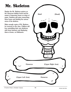


© Copyright 2025