
Cardiac involvement of primary hyperoxaluria accompanied by non
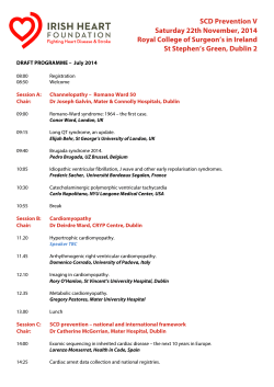
Türk Kardiyol Dern Arş - Arch Turk Soc Cardiol 2015;43(3):288-291 doi: 10.5543/tkda.2015.46026
288
Cardiac involvement of primary hyperoxaluria accompanied
by non-compaction cardiomyopathy and patent ductus arteriosus
Kalp tutulumu olan primer hiperoksalürili bir olguda
patent duktus arteriyozusun eşlik ettiği non kompaksiyon kardiyomiyopati
Nurcan Arat, M.D., Murat Akyıldız, M.D.,# Gürkan Tellioğlu, M.D.,* Yaman Tokat, M.D.†
Department of Cardiology, Istanbul Bilim University Faculty of Medicine, Istanbul
#
Department of Gastroenterology, Istanbul Bilim University Faculty of Medicine, Istanbul
*Department of Renal Transplantation, Sisli Florence Nightingale Hospital, Istanbul
†
Department of Liver Transplantation, Sisli Florence Nightingale Hospital, İstanbul
Summary– Primary hyperoxaluria is a rare hereditary metabolic disorder resulting in accumulation of calcium oxalate in
visceral organs, including the heart. We report a 19-year-old
male with non- compaction cardiomyopathy combined with
patent ductus arteriosus awaiting combined liver-kidney
transplantation for primary hyperoxaluria. After surgical closure of the patent ductus arteriosus, the patient underwent
a successful renal and subsequent liver transplantation.
The presence of hypertrophic cardiomyopathy in hyperoxaluria patients has been reported before, but this is the first
report of non-compaction myocardium with patent ductus
arteriosus in a patient with primary hyperoxaluria. At the
third month after combined liver and renal transplantation,
improvement in cardiac functions were observed. Primary
hyperoxaluria is a clinical entity to be taken into consideration in differential diagnosis of hypertrophied myocardium
with high myocardial echocardiographic intensity. In cases
of hyperoxaluria, additional congenital abnormalities may
complicate the clinical picture.
P
rimary hyperoxaluria is a rare hereditary metabolic disorder that leads to accumulation of calcium
oxalate in visceral organs. Although clinical symptoms are often associated with the kidney, calcium
oxalate can accumulate in all tissues, including the
heart.[1,2] Here, for the first time we report a case of
Özet– Primer hiperoksalüri kalp de dahil olmak üzere iç
organlarda, kalsiyum oksalat birikimi ile sonuçlanan nadir
görülen kalıtsal metabolik bir hastalıktır. Bu yazıda, primer
hiperoksalüri nedeniyle kombine karaciğer ve böbrek nakli planlanan 19 yaşında erkek hastada, non kompaksiyon
kardiyomiyopatiyle birlikte patent duktus arteriyozusun eşlik
ettiği hiperoksalürinin kalp tutulumu sunuldu. Patent duktus
arteriyozus cerrahi olarak kapatılarak hastaya önce böbrek
ve takiben karaciğer nakli başarıyla uygulandı. Hiperoksalüri ile birlikte hipertrofik kardiyomiyopati daha önce bildirilmiştir, ancak bizim olgumuzdaki gibi patent duktus arteriyozus ve non kompaksiyon kardiyomiyopati varlığı primer
hiperoksalürili hastalarda bu olguyla ilk kez bildirilmektedir.
Kalp tutulumu olan hiperoksalürili olguda kombine karaciğer
ve böbrek nakli sonrasında üçüncü ayda kalp fonksiyonlarında iyileşme gözlendi. Primer hiperoksalüri, miyokartta
artmış eko yoğunluğunun eşlik ettiği ventrikül hipertrofisinin
ekokardiyografik ayırıcı tanısında dikkate alınması gereken
bir klinik tablodur. Hiperoksalürili olgularda doğuştan gelen
ek anomaliler klinik durumu zorlaştırabilir.
cardiac involvement; a non- Abbreviation:
compacted cardiomyopathy PDA Patent ductus arteriosus
associated with patent ductus arteriosus (PDA) in a patient with primary hyperoxaluria. The echocardiographic findings are presented.
Received: October 03, 2014 Accepted: February 02, 2015
Correspondence: Dr. Nurcan Arat. İstanbul Cad., Zenginbahçe Konutları 1, Göktürk, Eyüp, 34075 İstanbul.
Tel: +90 212 - 212 88 11 e-mail: aratnurcan@gmail.com
© 2015 Turkish Society of Cardiology
Non-compaction cardiomyopathy and patent ductus arteriosus
CASE REPORT
A 19-year-old male patient with a diagnosis of primary hyperoxaluria and evaluation for liver-kidney
transplantation was referred to the cardiology clinic for preoperative cardiac evaluation. His history
showed that he had developed progressive renal failure secondary to nephrolithiasis, and 5 years previously had undergone a left nephrectomy. Hemodialysis had started 2 years prior to admission. The patient
had no cardiac symptoms other than fatigue. He had a
family history of consanguineous parents, and hyperoxaluria was diagnosed in his two siblings on family
screening.
Physical examination revealed blood pressure of
100/60 mmHg and heart rate of 72/min. Heart sounds
were diminished and there was a continuous 3rd degree murmur with spreading to the back of the pulmonary area. A pansystolic 2/6 degree murmur on mitral
and tricuspid focus was present. Electrocardiography
revealed sinus rhythm (Fig 1).
Transthoracic echocardiography revealed increased granular echo density on the interventricular
septum (Fig. 2a). Compacted and non-compacted
spongy trabecular meshwork, more pronounced in
the left ventricular apical region, was detected (Fig.
2b). There was mild enlargement of left ventricular
diameter and left ventricular systolic function was
mildly impaired (EF 47%). The right ventricle was
slightly hypertrophied and enlarged. There was mild
pericardial effusion and both atria were significantly
289
enlarged. Doppler echocardiography showed severe
tricuspid regurgitation and increased pulmonary artery systolic pressure (42 mmHg). Doppler echocardiography was also consistent with impaired
diastolic filling pattern. A large PDA and moderate
left-to-right shunt was detected by echocardiography
(Fig. 2c), and confirmed by cardiac catheterization
(Fig. 2d). Percutaneous closure of the PDA was not
possible because of the anatomy of the ductus, so it
was completely closed surgically. The patient underwent successful kidney and liver transplantation four
months after the cardiac operation. The pericardial
effusion disappeared and improvement in cardiac
diameters and systolic function (left ventricular end
systolic diameter decreased from 4.9 cm to 4.2 cm
and EF increased from 47% to 58%) was observed
on echocardiographic evaluation at the 3rd postoperative month. Echocardiographic screening of his two
asymptomatic hyperoxaluric siblings revealed only
an increase in myocardial echo density in a granular
manner, and no apparent structural or functional cardiac abnormalities were found.
DISCUSSION
Primary hyperoxaluria is a rare autosomal-recessive
disorder characterized by increased urinary excretion of calcium oxalate, recurrent urolithiasis, nephrocalcinosis, and accumulation of insoluble oxalate
throughout the body (oxalosis).[3-5] Patients are initially diagnosed with renal failure and subsequently have
the symptoms and signs of oxalosis. Major sites of deposition include bone, bone marrow, heart, blood vessels, joints, male urogenital system, central nervous
system, peripheral nerves, skin and soft tissues. Most
patients die as a result of uremia before or during the
third decade.
Even in industrialized countries, there is a high
rate of late diagnosis in advanced renal failure or after
kidney graft failure in the setting of isolated kidney
transplantation (up to 40% in adults), which denotes
underreporting.[6]
In severe cases, or in order to correct liver and kidney damage, combined liver-kidney transplantation to
correct the enzyme deficiency is the only viable alternative.[5]
Figure 1. Twelve-lead ECG recording shows sinus rhythm
with left axis deviation and non-specific ST-T wave changes
in D1, aVL leads.
In hyperoxaluria patients who have limited survival, cardiac abnormalities such as sudden arrhythmias
and heart blocks,[7] coronary and peripheral vascular
Türk Kardiyol Dern Arş
290
A
C
B
D
Figure 2. (A) Two-dimensional transthoracic echocardiography shows severe myocardial hypertrophy with granular, dense
echo in inter ventricular septum (arrows) and an enlarged left atrium (LA) and left ventricle (LV) on the left ventricular apical
3-chamber view. (B) Transthoracic color Doppler echocardiography shows the flow observed in the non-compacted myocardial
walls (arrows) in left ventricular (LV) apical segments on apical modified long-axis view. (C) Color M-mode echocardiography in
suprasternal view shows the continuous flow in the PDA (arrow) at the junction of the descending aorta. (D) PDA (arrow) shown
in aortography.
involvement,[1,8] atrial mass, increase in left ventricular mass index, hypertrophic cardiomyopathy,[9] left
atrial enlargement, pulmonary hypertension and diastolic dysfunction, decreased ejection fraction, infiltrative status and valve pathologies and right ventricular function deterioration have all been previously
reported.
However, PDA and non-compaction cardiomyopathy in a patient with hyperoxaluria have not been
encountered in the current literature. There are several
reports indicating improvement in cardiac function in
patients with hyperoxaluria after a combined liver
and kidney transplantation, as occurred in our case.
[2,10]
Echocardiographic examination at the 3rd postoperative month showed that left ventricular ejection
fraction had increased to 62%, pericardial effusion
had disappeared and pulmonary artery pressure had
decreased. Pericardial effusion and left ventricular
systolic functional improvement is thought to be due
to the impact of the actual volume overload change
as a result of elimination of both the PDA and renal
failure.
Primary hyperoxaluria is a clinical condition which
should be considered in the differential diagnosis of
ventricular hypertrophy accompanied by increased
echo density on echocardiography. The clinical picture and differential diagnosis can be complicated in a
patient with a combination of such congenital anomalies and hyperoxaluria.
Conflict-of-interest issues regarding the authorship or
article: None declared.
Non-compaction cardiomyopathy and patent ductus arteriosus
REFERENCES
1. Bogle MA, Teller CF, Tschen JA, Smith CA, Wang A. Primary hyperoxaluria in a 27-year-old woman. J Am Acad Dermatol 2003;49:725-8. CrossRef
2.Cochat P. Primary hyperoxaluria type 1. Kidney Int
1999;55:2533-47. CrossRef
3. Cochat P, Deloraine A, Rotily M, Olive F, Liponski I, Deries N. Epidemiology of primary hyperoxaluria type 1. Société
de Néphrologie and the Société de Néphrologie Pédiatrique.
Nephrol Dial Transplant 1995;10 Suppl 8:3-7. CrossRef
4. Danpure CJ, Jennings PR, Penketh RJ, Wise PJ, Cooper PJ,
Rodeck CH. Fetal liver alanine: glyoxylate aminotransferase
and the prenatal diagnosis of primary hyperoxaluria type 1.
Prenat Diagn 1989;9:271-81. CrossRef
5. Kemper MJ. Concurrent or sequential liver and kidney transplantation in children with primary hyperoxaluria type 1? Pediatr Transplant 2005;9:693-6. CrossRef
6. Hoppe B, Langman CB. A United States survey on diagnosis, treatment, and outcome of primary hyperoxaluria. Pediatr
Nephrol 2003;18:986-91. CrossRef
291
7. Quan KJ, Biblo LA. Type I primary hyperoxaluria: an unusual presentation of ventricular tachycardia. Cardiol Rev
2003;11:318-9. CrossRef
8. Marconi V, Mofid MZ, McCall C, Eckman I, Nousari HC. Primary hyperoxaluria: report of a patient with livedo reticularis
and digital infarcts. J Am Acad Dermatol 2002;46(2 Suppl
Case Reports):16-8.
9. Mookadam F, Smith T, Jiamsripong P, Moustafa SE, Monico
CG, Lieske JC, et al. Cardiac abnormalities in primary hyperoxaluria. Circ J 2010;74:2403-9. CrossRef
10.McDonald JC, Landreneau MD, Rohr MS, DeVault GA Jr.
Reversal by liver transplantation of the complications of primary hyperoxaluria as well as the metabolic defect. N Engl J
Med 1989;321:1100-3. CrossRef
Key words: Congenital abnormalities; ductus arteriosus, patent; hyperoxaluria, primary; kidney transplantation; liver transplantation.
Anahtar sözcükler: Doğumsal anomaliler; patent duktus arteriyozus; hiperoksalüri, birincil; böbrek transplantasyonu; karaciğer
transplantasyonu.


© Copyright 2025