L-4 Radiologic Investigation of Chest & CVS Diseases
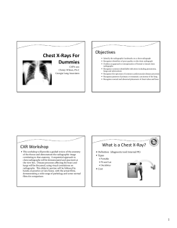
Done By: Sahar Alharthi COLOR GUIDE: • Females' Notes Reviewed By: Basil AlSuwaine • Males' Notes • Important • Additional • 431 team Radiologic Investigation of Chest & CVS Diseases 432RadiologyTeam Objectives Not Given ;( 1 Radiologic Investigation of Chest & CVS Diseases 432RadiologyTeam From Slides 431 Teams Extra All related work of 431 Teams is included here The role of chest x-ray in cardiac assessment is very limited, but the main role of chest x-ray in cardiac assessment is to detect the effect of cardiac conditions on the lungs, parenchyma & pulmonary vessels. • In normal adult, 2/3 of the heart is located in the left side whereas 1/3 of the heart is in the right side. • In children, 1/2 of the heart is in the left and the other 1/2 in the right side. • Otherwise than that, it will indicate abnormality. • Left cardiac contour is mainly formed by Left Ventricle & Aortic Knob (knuckle). • Right cardiac contour is mainly formed by Right Atrium & Ascending Aorta. The top 5 are the most important in making a diagnosis Left Side Dextrocardia is a condition in which the heart is located in the right side. Situs inversus: is a rare congenital condition in which the major visceral organs are reversed or mirrored from their normal positions. 2 = Lateral View Radiologic Investigation of Chest & CVS Diseases 432RadiologyTeam Normally, the vertebra is mild kyphosis (anterioposterior kyphosis) (S shaped). Any deviation of vertebra to the right or left is scoliosis. Here we have a scoliosis deviated to the left. (You have to mention in which side the deviation is) The heart is also deviated → to insure the reason of its deviation we do lateral CXR. Lateral view CXR reveals that this patient is also having a Pectus Exavatum → and it is the actual cause of heart deviation in addition to the scoliosis. Pectus Exavatum: is the most common congenital deformity of the anterior wall of the chest, in which several ribs and the sternum grow abnormally “depressed sternum”. This produces a caved-in or sunken appearance of the chest. 3 Radiologic Investigation of Chest & CVS Diseases 432RadiologyTeam The fissures are not seen in normal xrays (only 30% of pt) If we know the location of each fissure we can also know which lobe is involved (in case of any lung diseases). Always COMPARE both lungs looking for an abnormality. On the PA chest x-ray, the transverse (A) fissure divides the right middle lobe from the right upper lobe and is something not well seen. There is no Transverse fissure on the left. The oblique fissures (B) are usually not well seen on the PA view because you are looking through them obliquely. If there is fluid in the fissure, it is occasionally manifested as a density at the lower lateral margin. Normal Abnormal (Pleural effusion) The basic diagnostic instance is to detect an abnormality. In both cases, there is an abnormal opacity in the left upper lobe. The main difference is in the edges. It is important to differentiate between the two for the differential diagnosis. In the case ABOVE, the opacity would be best described as a mass, because its EDGES are well defined 3-D STRUCTURE. (Could be a tumor or abscess). The case BELOW, the opacity is poorly defined diffuse infiltration. Seen in airspace diseases such as pneumonia. (Most of the time with inflammatory diseases - like pneumonia - , but sometimes it can be seen in a tumor). Radiologists also depend on clinical data. E.g. if the 2nd image was for an old patient with no fever, neoplasm would be suspected. Pneumonia wouldn’t be as much, because fever is not a common symptom in older adults. http://www.webmd.com/lung/tc/pneumonia-symptoms 4 Radiologic Investigation of Chest & CVS Diseases 432RadiologyTeam Solitary = Single Nodule: opacity with demarcated margins (3D dimension) A solitary nodule in the lung can be totally innocuous or potentially a fatal lung cancer. After detection the initial step in analysis is to compare the film with prior films if available. A nodule that is unchanged for 2 years is almost certainly benign. Be sure to evaluate for the presence of multiple nodules, as this finding would change the differential entirely. If the nodule is indeterminate after considering old films and calcification, subsequent steps in the work-up include ordering a CT and a tissue biopsy. CT showing a mass that is likely pleural based (red arrows). Note the pleural effusion posteriorly. CT showing bone destruction indicative of an extrapleural mass. Localized Fibrous Tumor of the Pleura. Large, pleural-based soft tissue mass abuts. pleura in right lower lung with an acute angle (white arrow) and obtuse angle (yellow arrow) where it meets the chest wall. 5 Mediastinal windows show heterogeneous nature of the contrastenhancing mass with some areas of lower attenuation (blue arrows) most likely representing necrosis. Radiologic Investigation of Chest & CVS Diseases 432RadiologyTeam Loss of volume of lobe, segment or sub segment of the lung. Example: Collapse (lung), Atelectasis of the upper lobe, Atelectasis of the right lung, & Sub segmental atelectasis. Most common causes: Bronchial obstruction, Pneumothorax, & Pleural effusion. Loss of air in lobe, segment or sub segment of the lung. Example: Pneumonia (lobe), Tumor, TB, HF, Infections. Other common causes: Infarction, Contusion, & Immunological Disorders. Loss of air → what usually looks black will be white. Volume Loss Associated with ipsilateral shift Linear, wedge-shaped Apex at hilum Normal or ↑ Volume No shift, or if present then contralateral Consolidation, air space process Not centered at hilum Question asked: even with loss of volume it will look white, right? Doctor answered: I understand your question ;p , you mean in loss of volume of collapse there’s loss of air too. Yes, but the most important is loss of volume. Sometimes, you hear people saying “consolidation collapse”, what does that mean? It means the lung became white because of loss of air but also its volume became smaller. Therefore, the rule is (anything that looks white in the lung without any loss of volume will be called consolidation. And anything that looks white with loss of volume will be called atelectasis). When there’s loss of volume in an area, the structures near this area will move. As we’ve seen in upper lobe collapse, the fissure moved up. So, everything related to this lobe will move. If the area that lost volume was near the trachea then trachea will move, if it’s near the fissure then fissure will move. So, it is associated with ipsilateral move or shift. Most of pneumonias are normal in size. But if happened to be loss of volume in pneumonia then we call it (consolidation collapse). 6 Radiologic Investigation of Chest & CVS Diseases 432RadiologyTeam ↑ Density in the right middle lobe. No shift of transverse & oblique fissures (red arrows). Trachea is slightly moved to the other side. ↑ Density without loss of volume = Consolidation (Pneumonia) We saw something white on the right side, so we did a lateral view to investigate more. On lateral view, we found the same. Triangular opacity is associated with collapse. Green lines: normal fissures. Red lines: actual fissures as appeared in x-ray. The transverse fissure moved down, and the oblique fissure moved up. Shift of fissures → Loss of volume → Atelectasis ↑ Opacity (density) in the right lower lobe. PA view shows ↑ Density in the right (upper & middle) lobes. (Above the transverse fissure). Lateral view: normally, the oblique fissure is not seen, but here it is seen because the color is white. Transverse fissure is not seen. No volume loss → no shift of fissures → Consolidation (Pneumonia). There is ↑ density (opacity) in the right upper lobe. Transverse fissure moved upward (red arrows). Increased density + Shift of fissures = Loss of volume → Atelectasis of the right upper lobe. 7 Radiologic Investigation of Chest & CVS Diseases 432RadiologyTeam Alveolar spaces filled with … something Radiologist’s Report: “Consolidation” “Air space opacity” Fluffy density” Infiltrate” Non-specific: Atelectasis Pneumonia Bleeding Edema Tumor This Page was skipped by the doctor In radiology, the silhouette sign refers to the loss of normal borders between thoracic structures. A) Normal chest radiograph B) Q fever pneumonia affecting the right lower and middle lobes. Note the loss of the normal radiographic silhouette (contour) between the affected lung and its right heart border as well as between the affected lung and its right diaphragm border. This phenomenon is called “silhouette sign”. If an intrathoracic opacity is in anatomic contact with, for example, the heart border, then the opacity will obscure that border. The location of this abnormality can help to determine the location anatomically. Best sign – Shift of a fissure Rapid development & clearance Air bronchograms if non-obstructive Secondary signs: o Mediastinal shift o Elevated diaphragm o Ribs closer together o Vague increased density 8 Test Yourself: https://www.meded.virginia.edu/courses/rad/cxr/postquestions/posttest.ht ml https://www.meded.virginia.edu/courses/rad/cxr/introductionchest.html http://radiologymasterclass.co.uk/tutorials/chest/chest_pat hology/chest_pathology_start.html http://emedicine.medscape.com/article/296468workup#a0720 Radiologic Investigation of Chest & CVS Diseases 432RadiologyTeam The transverse fissure got back to its normal place. The patient had a tube that went too deep and closed the bronchus, so after the tube was removed; the patient got better within the next day. The transverse fissure is shifted upward (red arrows) because of upper lobe collapse in the right lung. Right Upper Lobe Atelectasis (RUL Atx). PA view shows ↑ density in the right middle lobe. Lateral view: there is shifting in both transverse and oblique fissures which indicate loss of volume of the middle lobe → Right Middle Lobe Atelectasis (RML Atx). Doctor said: There’s consolidation by loss of air & there’s atelectasis by loss of volume, so if you want to say “Consolidation Collapse” then it’s acceptable. And if you want to say “Collapse” only, it’s also accepted. But if you say “Consolidation” only, then it’s wrong. Because it’s make a different in the treatment. Remember that atelectasis takes the upper hand so if it exists you have to mention it. 9 432RadiologyTeam Radiologic Investigation of Chest & CVS Diseases PA view shows an abnormal left lung with no displacement of the trachea. Lateral view: The oblique fissure (red line) is moved upwards towards the collapsed side (red arrows). Left Upper Lobe Atelectasis (LUL Atx). Skipped by the doctor as he said it confuses a lot of people. Left Lower Lobe Collapse In atelectasis, the trachea & the heart are deviated toward the affected side. http://emedicine.medscape.com/article/296468-clinical#a0217 An example of right tension pneumothorax, the mediastinum is displaced to the left. (Step-Up to Medicine. 3rd edition. page 87) So we ask ourselves: 1. Is there a white color (density)? If yes, then we have a diffuse lung disease. 2. Is there a loss of volume? o If yes, then atelectasis (collapse). o If not, then it’s consolidation (pneumonia). 11 432RadiologyTeam Radiologic Investigation of Chest & CVS Diseases Signs: Air bronchogram Silhouette – “positive” or “negative” Dense hilum “spine” sign All are signs of any air space process Dx of pneumonia depends on appropriate clinical scenario. One of the most important signs that indicate consolidation is “Air Bronchogram”. Now, if we want to see the bronchioles, we have 2 choices: 1. The lung color will be all white, so I can see the bronchiole. (Because normally it’s all black in black and I can’t see the bronchioles). 2. We do what used to be called an air bronchogram technique, which consists of injecting a contrast in the bronchioles that makes it look white in the x-ray. (Bronchioles will look white). Of course this all is in the past, now we do high resolution CT, which can show us everything or hide somethings and show others; meaning that we can see bronchioles easily thanks to god. So, air bronchogram sign means that when there’s consolidation, the lung will be white of course, and bronchioles are already black; and so we can see bronchioles as black lines. 11 Radiologic Investigation of Chest & CVS Diseases 432RadiologyTeam Air bronchogram Blood vessels Pneumonia Normal Air bronchogram is important because if you have consolidation like this with no air bronchogram, it means that the bronchial tree is obstructed by a tumor. You can see bronchial tree black in color d/t surrounding whitish diseased lung parenchyma. Ok, so why do we say consolidation or collapse? Simply, one of the most important reasons in children that cause lung collapse is foreign body. So, if a child came with a lung collapse and I diagnosed him with a consolidation and I gave him a medical treatment, then he will never get better because the foreign body is still there. If it’s an elderly, we look for something that may obstruct the bronchus which will cause the collapse. For example, lymph nodes, lung mass, or tumor in the bronchus. 12 432RadiologyTeam Radiologic Investigation of Chest & CVS Diseases Right Middle Lobe Right Upper Lobe Right Lower Lobe 13 432RadiologyTeam Radiologic Investigation of Chest & CVS Diseases Compare Costo-Phrenic Angles The Costo-Phrenic angle is the most dependent part of the pleural cavity. If there’s a collection of water, it will be blocked. Pleural effusion is presence of fluids in the pleural cavity. On an upright film, an effusion will cause blunting on the lateral and if large enough, the posterior costophrenic sulci. Sometimes a depression of the involved diaphragm will occur. A large effusion can lead to a mediastinal shift away from the effusion and opacity the hemithorax. Approximately 200 ml of fluid are needed to detect an effusion in the frontal film vs. approximately 75ml for the lateral. Larger effusions, especially if unilateral, are more likely to be caused by malignancy than smaller ones. The Example shows PA and lateral film of a patient with bilateral pleural effusions. (Note the concave menisci blunting both posterior costophrenic angles). The CT above on the left demonstrates a large infected pleural fluid collection, an empyema. Compare and contrast that to the image on the right which is an intrapulmonary abscess. 14 432RadiologyTeam Radiologic Investigation of Chest & CVS Diseases A pneumothorax is defined as air inside the thoracic cavity but outside the lung. A spontaneous pneumothorax (PTX) is one that occurs without an obvious inciting incident. Some causes of spontaneous PTX are; idiopathic, asthma, COPD, pulmonary infection, neoplasm, Marfanâs syndrome, and smoking cocaine. Picture A: shows pneumomediastinum Picture B: This image shows a close-up of a pneumothorax in an upright PA film as a white pleural line (red arrow) with atmospheric air outside of it. No pulmonary vascular markings are seen outside of the line. Notice the predilection to the apices and the periphery. For further explanation of Pneumothorax: https://www.meded.virginia.edu/courses/rad/cxr/pathology8chest.html 15 432RadiologyTeam Radiologic Investigation of Chest & CVS Diseases CT scans clearly demonstrating the presence of air in the mediastinum (red arrows) and subcutaneous emphysema (yellow arrows). Findings for pneumomediastinum include; streaky lucencies over the mediastinum that extend into the neck, and elevation of the parietal pleura along the mediastinal borders. Causes of pneumomediastinum include; asthma, surgery (post-op complication), traumatic tracheobronchial rupture, abrupt changes in intrathoracic pressure (vomiting, coughing, exercise, parturition), ruptured esophagus, barotrauma, and smoking crack cocaine. For further explanation of Pneumomediastinum: https://www.med-ed.virginia.edu/courses/rad/cxr/pathology15chest.html The above three images show a hydropneumothorax in three different views. The PA, lateral, and right decube reveal a layering out of the air and fluid. The right decube film demonstrates a right hydropneumothorax. (Note the pleural air/fluid level demonstrated by the horizontal air/fluid interface (arrows)). CT chest: Arrow A- air, B- Fluid. Large hydropneumothorax, unilocular, some pleural thickening. Appearance suggestive of empyema. Associated collapse of right lung. 16 432RadiologyTeam Radiologic Investigation of Chest & CVS Diseases Emphysema is loss of elastic recoil of the lung with destruction of pulmonary capillary bed and alveolar septa. It is caused most often by cigarette smoking and less commonly by alpha-1 antitrypsin deficiency. Emphysema is commonly seen on CXR as diffuse hyperinflation with flattening of diaphragms, increased retrosternal space, bullae (lucent, air-containing spaces that have no vessels that are not perfused) and enlargement of PA/RV (secondary to chronic hypoxia) an entity also known as cor pulmonale. Hyperinflation and bullae are the best radiographic predictors of emphysema. For Further explanation of Emphysema: https://www.med-ed.virginia.edu/courses/rad/cxr/pathology10chest.html 17 432RadiologyTeam Radiologic Investigation of Chest & CVS Diseases SUMMARY 1. Pectus Exavatum: is the most common congenital deformity of the anterior wall of the chest, in which several ribs and the sternum grow abnormally “depressed sternum”. 2. Dextrocardia: is a condition in which the heart is located in the right side. 3. A well-defined opacity (3D structure) is usually a mass. 4. Poorly defined opacity is diffuse infiltration. 5. Atelectasis (collapse) → loss of volume. 6. Consolidation (pneumonia) → loss of air. (air bronchogram) 7. Air bronchograms can occur in both collapse and pneumonia. 8. Atelectasis best sign is shift of fissures. 9. Remember loss of volume (atelectasis) always takes the upper hand; meaning when it exists it has to be mentioned. 10. Most important reason of collapse in elderly is Lung Tumor. 11. Most important reason of collapse in children is Foreign Body. 12. Pleural effusion is presence of fluids in the pleural cavity. 13. A pneumothorax is defined as air inside the thoracic cavity but outside the lung. 14. A hydropneumothorax is both air and fluid in the pleural space. 15. Emphysema is loss of elastic recoil of the lung with destruction of pulmonary capillary bed and alveolar septa. 16. Hyperinflation and bullae are the best radiographic predictors of emphysema. 18 Radiologic Investigation of Chest & CVS Diseases 432RadiologyTeam Questions 1) Patient is administered to the hospital with fever and cough, CT was done and showed? a. b. c. d. 2) Pulmonary embolism Pneumonia Right lung mass Right lung collapse Identify the abnormality shown in the image below? a. b. c. d. Right middle lobe pneumonia Right upper lobe pneumonia Right lower lobe pneumonia Right upper lobe atelectasis 432 Radiology Team Leaders Answers: 1st Questions: B Eman AlBedaie Emansaleh202 @gmail.com 2nd Questions: B Basil AlSuwaine Basil.als@hotmail.com 19
© Copyright 2025


